1W9X | pdb_00001w9x

Bacillus halmapalus alpha amylase


Experimental Data Snapshot

  • Method: X-RAY DIFFRACTION
  • Resolution: 2.10 Å
  • R-Value Free: 
    0.198 (Depositor) 
  • R-Value Work: 
    0.151 (Depositor), 0.154 (DCC) 
  • R-Value Observed: 
    0.152 (Depositor) 

Starting Model: experimental
View more details

wwPDB Validation 3D Report Full Report

Validation slider image for 1W9X

This is version 2.1 of the entry. See complete history

Literature

Structure of a Bacillus Halmapalus Family 13 Alpha-Amylase, Bha, in Complex with an Acarbose-Derived Nonasaccharide at 2.1 A Resolution

Davies, G.J.Brzozowski, A.M.Dauter, Z.Rasmussen, M.D.Borchert, T.V.Wilson, K.S.

(2005) Acta Crystallogr D Biol Crystallogr 61: 190

  • DOI: https://doi.org/10.1107/S0907444904027118
  • Primary Citation Related Structures: 
    1W9X

  • PubMed Abstract: 

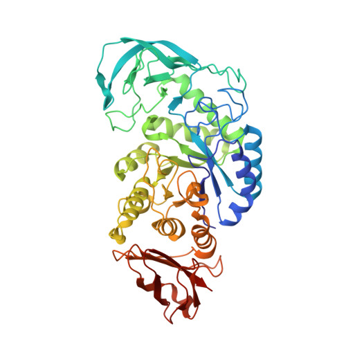
    The enzymatic digestion of starch by alpha-amylases is one of the key biotechnological reactions of recent times. In the search for industrial biocatalysts, the family GH13 alpha-amylase BHA from Bacillus halmapalus has been cloned and expressed. The three-dimensional structure at 2.1 A resolution has been determined in complex with the (pseudo)tetrasaccharide inhibitor acarbose. Acarbose is found bound as a nonasaccharide transglycosylation product spanning the -6 to +3 subsites. Careful inspection of electron density suggests that the bound ligand could not have been formed through successive transglycosylations of acarbose and must also have featured maltose or maltooligosaccharides as an acceptor.


  • Organizational Affiliation
    • Department of Chemistry, University of York, England. davies@ysbl.york.ac.uk

Macromolecule Content 

  • Total Structure Weight: 56.86 kDa 
  • Atom Count: 4,300 
  • Modeled Residue Count: 481 
  • Deposited Residue Count: 481 
  • Unique protein chains: 1

Macromolecules

Find similar proteins by:|  3D Structure
Entity ID: 1
MoleculeChains  Sequence LengthOrganismDetailsImage
ALPHA AMYLASE481Sutcliffiella halmapalaMutation(s): 0 
EC: 3.2.1.1
UniProt
Find proteins for A0ACD6B935 (Bacillus sp)
Explore A0ACD6B935 
Go to UniProtKB:  A0ACD6B935
Entity Groups
Sequence Clusters30% Identity50% Identity70% Identity90% Identity95% Identity100% Identity
UniProt GroupA0ACD6B935
Sequence Annotations
Expand
Reference Sequence

Oligosaccharides

Help  
Entity ID: 2
MoleculeChains Length2D Diagram GlycosylationD Interactions
4,6-dideoxy-4-{[(1S,4R,5S,6S)-4,5,6-trihydroxy-3-(hydroxymethyl)cyclohex-2-en-1-yl]amino}-alpha-D-glucopyranose-(1-4)-alpha-D-glucopyranose-(1-4)-alpha-D-glucopyranose-(1-4)-alpha-D-glucopyranose-(1-4)-4,6-dideoxy-4-{[(1S,4R,5S,6S)-4,5,6-trihydroxy-3-(hydroxymethyl)cyclohex-2-en-1-yl]amino}-alpha-D-glucopyranose-(1-4)-alpha-D-glucopyranose-(1-4)-beta-D-glucopyranose
B
7N/AN/A
Glycosylation Resources
GlyTouCan: G14192NX
GlyCosmos: G14192NX

Experimental Data & Validation

Experimental Data

  • Method: X-RAY DIFFRACTION
  • Resolution: 2.10 Å
  • R-Value Free:  0.198 (Depositor) 
  • R-Value Work:  0.151 (Depositor), 0.154 (DCC) 
  • R-Value Observed: 0.152 (Depositor) 
Space Group: P 21 21 21
Unit Cell:
Length ( Å )Angle ( ˚ )
a = 48.18α = 90
b = 75.847β = 90
c = 155.23γ = 90
Software Package:
Software NamePurpose
REFMACrefinement
DENZOdata reduction
SCALEPACKdata scaling
AMoREphasing

Structure Validation

View Full Validation Report


Entry History 

Deposition Data

Revision History  (Full details and data files)

  • Version 1.0: 2005-02-09
    Type: Initial release
  • Version 1.1: 2011-05-08
    Changes: Version format compliance
  • Version 1.2: 2011-07-13
    Changes: Version format compliance
  • Version 2.0: 2020-07-29
    Type: Remediation
    Reason: Carbohydrate remediation
    Changes: Atomic model, Data collection, Derived calculations, Other, Structure summary
  • Version 2.1: 2023-12-13
    Changes: Data collection, Database references, Refinement description, Structure summary