1W74 | pdb_00001w74

X-ray structure of peptidyl-prolyl cis-trans isomerase A, PpiA, Rv0009, from Mycobacterium tuberculosis.


Experimental Data Snapshot

  • Method: X-RAY DIFFRACTION
  • Resolution: 2.60 Å
  • R-Value Free: 
    0.229 (Depositor), 0.237 (DCC) 
  • R-Value Work: 
    0.212 (Depositor), 0.221 (DCC) 
  • R-Value Observed: 
    0.213 (Depositor) 

Starting Model: experimental
View more details

wwPDB Validation 3D Report Full Report

Validation slider image for 1W74

This is version 1.3 of the entry. See complete history

Literature

X-Ray Structure of Peptidyl-Prolyl Cis-Trans Isomerase a from Mycobacterium Tuberculosis

Henriksson, L.M.Johansson, P.Unge, T.Mowbray, S.L.

(2004) Eur J Biochem 271: 4107

  • DOI: https://doi.org/10.1111/j.1432-1033.2004.04348.x
  • Primary Citation Related Structures: 
    1W74

  • PubMed Abstract: 

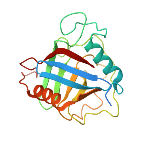
    Peptidyl-prolyl cis-trans isomerases (EC 5.2.1.8) catalyse the interconversion of cis and trans peptide bonds and are therefore considered to be important for protein folding. They are also thought to participate in processes such as signalling, cell surface recognition, chaperoning and heat-shock response. Here we report the soluble expression of recombinant Mycobacterium tuberculosis peptidyl-prolyl cis-trans isomerase PpiA in Escherichia coli, together with an investigation of its structure and biochemical properties. The protein was shown to be active in a spectrophotometric assay, with an estimated kcat/Km of 2.0 x 10(6) m(-1).s(-1). The X-ray structure of PpiA was solved by molecular replacement, and refined to a resolution of 2.6 A with R and Rfree values of 21.3% and 22.9%, respectively. Comparisons to known structures show that the PpiA represents a slight variation on the peptidyl-prolyl cis-trans isomerase fold, previously not represented in the Protein Data Bank. Inspection of the active site suggests that specificity for substrates and cyclosporin A will be similar to that found for most other enzymes of this structural family. Comparison to the sequence of the second M. tuberculosis enzyme, PpiB, suggests that binding of peptide substrates as well as cyclosporin A may differ in that case.


  • Organizational Affiliation
    • Department of Cell and Molecular Biology, Uppsala University, Sweden.

Macromolecule Content 

  • Total Structure Weight: 40.61 kDa 
  • Atom Count: 2,575 
  • Modeled Residue Count: 342 
  • Deposited Residue Count: 382 
  • Unique protein chains: 1

Macromolecules

Find similar proteins by:|  3D Structure
Entity ID: 1
MoleculeChains  Sequence LengthOrganismDetailsImage
PEPTIDYL-PROLYL CIS-TRANS ISOMERASE A
A, B
191Mycobacterium tuberculosis H37RvMutation(s): 0 
EC: 5.2.1.8
UniProt
Find proteins for P9WHW3 (Mycobacterium tuberculosis (strain ATCC 25618 / H37Rv))
Explore P9WHW3 
Go to UniProtKB:  P9WHW3
Entity Groups
Sequence Clusters30% Identity50% Identity70% Identity90% Identity95% Identity100% Identity
UniProt GroupP9WHW3
Sequence Annotations
Expand
Reference Sequence

Experimental Data & Validation

Experimental Data

  • Method: X-RAY DIFFRACTION
  • Resolution: 2.60 Å
  • R-Value Free:  0.229 (Depositor), 0.237 (DCC) 
  • R-Value Work:  0.212 (Depositor), 0.221 (DCC) 
  • R-Value Observed: 0.213 (Depositor) 
Space Group: P 31
Unit Cell:
Length ( Å )Angle ( ˚ )
a = 65.349α = 90
b = 65.349β = 90
c = 102.493γ = 120
Software Package:
Software NamePurpose
REFMACrefinement
MOSFLMdata reduction
SCALAdata scaling
AMoREphasing

Structure Validation

View Full Validation Report


Entry History 

Deposition Data

Revision History  (Full details and data files)

  • Version 1.0: 2004-10-20
    Type: Initial release
  • Version 1.1: 2011-05-08
    Changes: Version format compliance
  • Version 1.2: 2011-07-13
    Changes: Version format compliance
  • Version 1.3: 2023-12-13
    Changes: Data collection, Database references, Other, Refinement description