1VYX | pdb_00001vyx

Solution structure of the KSHV K3 N-terminal domain


Experimental Data Snapshot

  • Method: SOLUTION NMR
  • Conformers Calculated: 30 
  • Conformers Submitted: 28 
  • Selection Criteria: NO VIOLATIONS GREATER THAN 0.25A, NO ANGLE VIOLATIONS 

wwPDB Validation 3D Report Full Report

Validation slider image for 1VYX

This is version 1.5 of the entry. See complete history

Literature

Solution Structure of the Kaposi'S Sarcoma-Associated Herpesvirus K3 N-Terminal Domain Reveals a Novel E2-Binding C4Hc3-Type Ring Domain

Dodd, R.B.Allen, M.D.Brown, S.E.Sanderson, C.M.Duncan, L.M.Lehner, P.J.Bycroft, M.Read, R.J.

(2004) J Biological Chem 279: 53840

  • DOI: https://doi.org/10.1074/jbc.M409662200
  • Primary Citation Related Structures: 
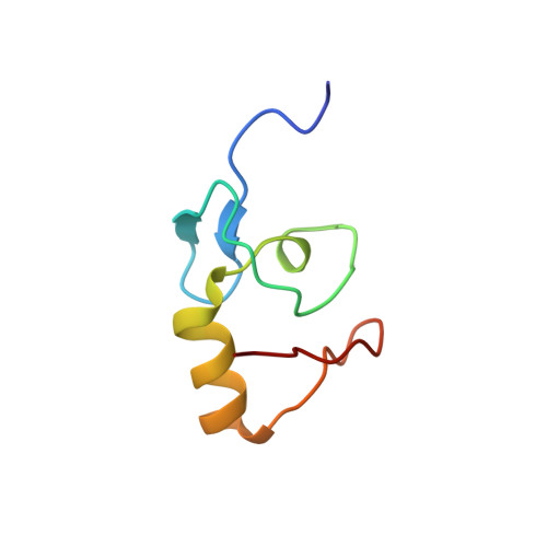
    1VYX

  • PubMed Abstract: 

    RING domains are found in a large number of eukaryotic proteins. Most function as E3 ubiquitin-protein ligases, catalyzing the terminal step in the ubiquitination process. Structurally, these domains have been characterized as binding two zinc ions in a stable cross-brace motif. The tumorigenic human gamma-herpesvirus Kaposi's sarcoma-associated herpesvirus encodes a ubiquitin-protein ligase termed K3, which functions as an immune evasion molecule by ubiquitinating major histocompatibility complex class I. K3 possesses at its N terminus a domain related to cellular RING domains but with an altered zinc ligand arrangement. This domain was initially characterized as a plant homeodomain, a structure not previously known to function as an E3. Here, it is conclusively demonstrated that the K3 N-terminal domain is a variant member of the RING domain family and not a plant homeodomain. The domain is found to interact with the cellular ubiquitin-conjugating enzymes UbcH5A to -C and UbcH13, which dock to the equivalent surface as on classical cellular RING domains. Interaction with UbcH13 suggests a possible role for K3 in catalyzing Lys(63)-linked ubiquitination.


  • Organizational Affiliation
    • Cambridge Institute for Medical Research, Wellcome Trust/MRC Building, Addenbrooke's Hospital, Hills Road, Cambridge CB2 2XY, UK.

Macromolecule Content 

  • Total Structure Weight: 6.96 kDa 
  • Atom Count: 474 
  • Modeled Residue Count: 60 
  • Deposited Residue Count: 60 
  • Unique protein chains: 1

Macromolecules

Find similar proteins by:|  3D Structure
Entity ID: 1
MoleculeChains  Sequence LengthOrganismDetailsImage
ORF K360Human gammaherpesvirus 8Mutation(s): 0 
EC: 2.3.2.36
UniProt
Find proteins for P90495 (Human herpesvirus 8 type P (isolate GK18))
Explore P90495 
Go to UniProtKB:  P90495
Entity Groups
Sequence Clusters30% Identity50% Identity70% Identity90% Identity95% Identity100% Identity
UniProt GroupP90495
Sequence Annotations
Expand
Reference Sequence

Small Molecules

Experimental Data & Validation

Experimental Data

  • Method: SOLUTION NMR
  • Conformers Calculated: 30 
  • Conformers Submitted: 28 
  • Selection Criteria: NO VIOLATIONS GREATER THAN 0.25A, NO ANGLE VIOLATIONS 

Structure Validation

View Full Validation Report


Entry History 

Deposition Data

Revision History  (Full details and data files)

  • Version 1.0: 2004-10-01
    Type: Initial release
  • Version 1.1: 2011-05-08
    Changes: Version format compliance
  • Version 1.2: 2011-07-13
    Changes: Version format compliance
  • Version 1.3: 2018-01-24
    Changes: Source and taxonomy
  • Version 1.4: 2018-05-02
    Changes: Data collection, Experimental preparation
  • Version 1.5: 2024-05-15
    Changes: Data collection, Database references, Derived calculations, Other