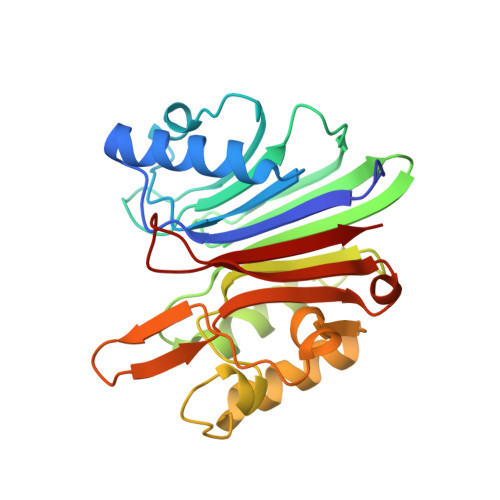
1VYB

Endonuclease domain of human LINE1 ORF2p


Experimental Data Snapshot

  • Method: X-RAY DIFFRACTION
  • Resolution: 1.80 Å
  • R-Value Free: 0.223 
  • R-Value Work: 0.186 
  • R-Value Observed: 0.188 

Starting Model: experimental
View more details

wwPDB Validation   3D Report Full Report


This is version 1.4 of the entry. See complete history


Literature

Crystal structure of the targeting endonuclease of the human LINE-1 retrotransposon.

Weichenrieder, O.Repanas, K.Perrakis, A.

(2004) Structure 12: 975-986

  • DOI: https://doi.org/10.1016/j.str.2004.04.011
  • Primary Citation of Related Structures:  
    1VYB

  • PubMed Abstract: 

    The human L1 endonuclease (L1-EN) is encoded by the non-LTR retrotransposon LINE-1 (L1). L1 is responsible for more than 1.5 million retrotransposition events in the history of the human genome, contributing more than a quarter to human genomic DNA (L1 and Alu elements). L1-EN is related to the well-understood human DNA repair endonuclease APE1, and its nicking specificity is a major determinant for retrotransposon integration site selection. The crystal structure of human L1 endonuclease is the first of a retrotransposon-encoded protein and a prototype for retrotransposon-encoded endonucleases involved in target-primed reverse transcription. Structure-based endonuclease alignments reveal a conserved threonine in addition to previously identified invariant residues and suggest that DNA recognition proceeds via the accommodation of an extrahelical nucleotide within a pocket of the enzyme. The present analysis will help to refine phylogenetic and functional relationships among metal-dependent phosphohydrolases and provides a basis for manipulating non-LTR retrotransposon integration site selection.


  • Organizational Affiliation

    The Netherlands Cancer Institute, Department of Molecular Carcinogenesis-H2, Plesmanlaan 121, 1066 CX Amsterdam, The Netherlands. o.weichenrieder@nki.nl


Macromolecules
Find similar proteins by:  (by identity cutoff)  |  3D Structure
Entity ID: 1
MoleculeChains Sequence LengthOrganismDetailsImage
ORF2 CONTAINS A REVERSE TRANSCRIPTASE DOMAIN
A, B
238Homo sapiensMutation(s): 0 
UniProt
Find proteins for O00370 (Homo sapiens)
Explore O00370 
Go to UniProtKB:  O00370
Entity Groups  
Sequence Clusters30% Identity50% Identity70% Identity90% Identity95% Identity100% Identity
UniProt GroupO00370
Sequence Annotations
Expand
  • Reference Sequence
Small Molecules
Ligands 3 Unique
IDChains Name / Formula / InChI Key2D Diagram3D Interactions
SO4
Query on SO4

Download Ideal Coordinates CCD File 
C [auth A]
D [auth A]
E [auth A]
H [auth B]
I [auth B]
C [auth A],
D [auth A],
E [auth A],
H [auth B],
I [auth B],
J [auth B],
K [auth B]
SULFATE ION
O4 S
QAOWNCQODCNURD-UHFFFAOYSA-L
GOL
Query on GOL

Download Ideal Coordinates CCD File 
G [auth A]GLYCEROL
C3 H8 O3
PEDCQBHIVMGVHV-UHFFFAOYSA-N
SO3
Query on SO3

Download Ideal Coordinates CCD File 
F [auth A],
L [auth B]
SULFITE ION
O3 S
LSNNMFCWUKXFEE-UHFFFAOYSA-L
Experimental Data & Validation

Experimental Data

  • Method: X-RAY DIFFRACTION
  • Resolution: 1.80 Å
  • R-Value Free: 0.223 
  • R-Value Work: 0.186 
  • R-Value Observed: 0.188 
  • Space Group: P 21 21 2
Unit Cell:
Length ( Å )Angle ( ˚ )
a = 91.044α = 90
b = 126.477β = 90
c = 43.004γ = 90
Software Package:
Software NamePurpose
REFMACrefinement
DENZOdata reduction
SCALEPACKdata scaling
AMoREphasing

Structure Validation

View Full Validation Report


Entry History 

Deposition Data

Revision History  (Full details and data files)

  • Version 1.0: 2004-06-04
    Type: Initial release
  • Version 1.1: 2011-05-08
    Changes: Version format compliance
  • Version 1.2: 2011-07-13
    Changes: Version format compliance
  • Version 1.3: 2018-02-28
    Changes: Database references, Source and taxonomy
  • Version 1.4: 2023-12-13
    Changes: Data collection, Database references, Other, Refinement description