1VTN | pdb_00001vtn

CO-CRYSTAL STRUCTURE OF THE HNF-3/FORK HEAD DNA-RECOGNITION MOTIF RESEMBLES HISTONE H5


Experimental Data Snapshot

  • Method: X-RAY DIFFRACTION
  • Resolution: 2.50 Å
  • R-Value Work: 
    0.211 (Depositor) 
  • R-Value Observed: 
    0.211 (Depositor) 

wwPDB Validation 3D Report Full Report

Validation slider image for 1VTN

This is version 1.2 of the entry. See complete history

Literature

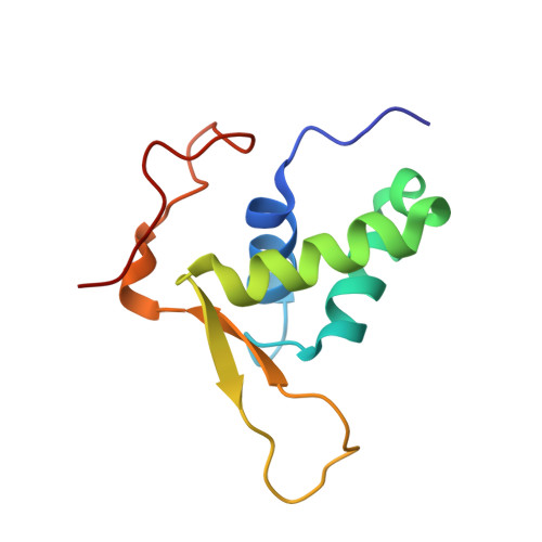
Co-Crystal Structure of the HNF-3/Fork Head DNA-Recognition Motif Resembles Histone H5

Clark, K.L.Halay, E.D.Lai, E.Burley, S.K.

(1993) Nature 364: 412-420

  • DOI: https://doi.org/10.1038/364412a0
  • Primary Citation Related Structures: 
    1VTN

  • PubMed Abstract: 

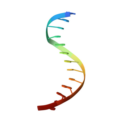
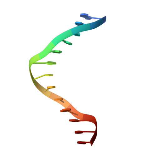
    The three-dimensional structure of an HNF-3/fork head DNA-recognition motif complexed with DNA has been determined by X-ray crystallography at 2.5 A resolution. This alpha/beta protein binds B-DNA as a monomer, through interactions with the DNA backbone and through both direct and water-mediated major and minor groove base contacts, inducing a 13 degrees bend. The transcription factor fold is very similar to the structure of histone H5. In its amino-terminal half, three alpha-helices adopt a compact structure that presents the third helix to the major groove. The remainder of the protein includes a twisted, antiparallel beta-structure and random coil that interacts with the minor groove.


  • Organizational Affiliation
    • Laboratory of Molecular Biophysics, Rockefeller University, New York, New York 10021.

Macromolecule Content 

  • Total Structure Weight: 20.02 kDa 
  • Atom Count: 1,381 
  • Modeled Residue Count: 128 
  • Deposited Residue Count: 128 
  • Unique protein chains: 1
  • Unique nucleic acid chains: 2

Macromolecules


Find similar proteins by:|  3D Structure
Entity ID: 3
MoleculeChains  Sequence LengthOrganismDetailsImage
HNF-3/FORK HEAD DNA-RECOGNITION MOTIF102Homo sapiensMutation(s): 0 
UniProt & NIH Common Fund Data Resources
Find proteins for P55318 (Homo sapiens)
Explore P55318 
Go to UniProtKB:  P55318
PHAROS:  P55318
GTEx:  ENSG00000170608 
Entity Groups
Sequence Clusters30% Identity50% Identity70% Identity90% Identity95% Identity100% Identity
UniProt GroupP55318
Sequence Annotations
Expand
Reference Sequence
Find similar nucleic acids by:  Sequence
Entity ID: 1
MoleculeChains LengthOrganismImage
DNA (5'-D(*GP*AP*CP*TP*AP*AP*GP*TP*CP*AP*AP*CP*C)-3')13N/A
Sequence Annotations
Expand
Reference Sequence
Find similar nucleic acids by:  Sequence
Entity ID: 2
MoleculeChains LengthOrganismImage
DNA (5'-D(*GP*GP*TP*TP*GP*AP*CP*TP*TP*AP*GP*TP*C)-3')13N/A
Sequence Annotations
Expand
Reference Sequence

Small Molecules

Ligands 1 Unique
IDChains Name / Formula / InChI Key2D Diagram3D Interactions
MG

Query on MG


Download:Ideal Coordinates CCD File
D [auth C]MAGNESIUM ION
Mg
JLVVSXFLKOJNIY-UHFFFAOYSA-N

Experimental Data & Validation

Experimental Data

  • Method: X-RAY DIFFRACTION
  • Resolution: 2.50 Å
  • R-Value Work:  0.211 (Depositor) 
  • R-Value Observed: 0.211 (Depositor) 
Space Group: P 31
Unit Cell:
Length ( Å )Angle ( ˚ )
a = 74.6α = 90
b = 74.6β = 90
c = 85.05γ = 120
Software Package:
Software NamePurpose
X-PLORrefinement

Structure Validation

View Full Validation Report


Entry History 

Deposition Data

Revision History  (Full details and data files)

  • Version 1.0: 2011-07-13
    Type: Initial release
  • Version 1.1: 2021-02-03
    Changes: Database references, Derived calculations, Structure summary
  • Version 1.2: 2023-12-27
    Changes: Data collection, Database references