1VGJ | pdb_00001vgj

Crystal structure of 2'-5' RNA ligase from Pyrococcus horikoshii


Experimental Data Snapshot

  • Method: X-RAY DIFFRACTION
  • Resolution: 1.94 Å
  • R-Value Free: 
    0.265 (Depositor), 0.268 (DCC) 
  • R-Value Work: 
    0.210 (Depositor), 0.220 (DCC) 
  • R-Value Observed: 
    0.216 (Depositor) 

Starting Model: experimental
View more details

wwPDB Validation 3D Report Full Report

Validation slider image for 1VGJ

This is version 1.5 of the entry. See complete history

Literature

The structure of Pyrococcus horikoshii 2'-5' RNA ligase at 1.94 A resolution reveals a possible open form with a wider active-site cleft

Gao, Y.G.Yao, M.Okada, A.Tanaka, I.

(2006) Acta Crystallogr Sect F Struct Biol Cryst Commun 62: 1196-1200

  • DOI: https://doi.org/10.1107/S1744309106046616
  • Primary Citation Related Structures: 
    1VGJ

  • PubMed Abstract: 

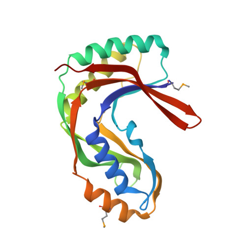
    Bacterial and archaeal 2'-5' RNA ligases, members of the 2H phosphoesterase superfamily, catalyze the linkage of the 5' and 3' exons via a 2'-5'-phosphodiester bond during tRNA-precursor splicing. The crystal structure of the 2'-5' RNA ligase PH0099 from Pyrococcus horikoshii OT3 was solved at 1.94 A resolution (PDB code 1vgj). The molecule has a bilobal alpha+beta arrangement with two antiparallel beta-sheets constituting a V-shaped active-site cleft, as found in other members of the 2H phosphoesterase superfamily. The present structure was significantly different from that determined previously at 2.4 A resolution (PDB code 1vdx) in the active-site cleft; the entrance to the cleft is wider and the active site is easily accessible to the substrate (RNA precursor) in our structure. Structural comparison with the 2'-5' RNA ligase from Thermus thermophilus HB8 also revealed differences in the RNA precursor-binding region. The structural differences in the active-site residues (tetrapeptide motifs H-X-T/S-X) between the members of the 2H phosphoesterase superfamily are discussed.


  • Organizational Affiliation
    • Faculty of Advanced Life Sciences, Graduate School of Life Sciences, Hokkaido University, Sapporo 060-0810, Japan.

Macromolecule Content 

  • Total Structure Weight: 21.27 kDa 
  • Atom Count: 1,529 
  • Modeled Residue Count: 184 
  • Deposited Residue Count: 184 
  • Unique protein chains: 1

Macromolecules

Find similar proteins by:|  3D Structure
Entity ID: 1
MoleculeChains  Sequence LengthOrganismDetailsImage
Hypothetical protein PH0099184Pyrococcus horikoshiiMutation(s): 3 
Gene Names: PH0099
EC: 3.1.4.58
UniProt
Find proteins for O57823 (Pyrococcus horikoshii (strain ATCC 700860 / DSM 12428 / JCM 9974 / NBRC 100139 / OT-3))
Explore O57823 
Go to UniProtKB:  O57823
Entity Groups
Sequence Clusters30% Identity50% Identity70% Identity90% Identity95% Identity100% Identity
UniProt GroupO57823
Sequence Annotations
Expand
Reference Sequence

Small Molecules

Modified Residues  1 Unique
IDChains TypeFormula2D DiagramParent
MSE
Query on MSE
A
L-PEPTIDE LINKINGC5 H11 N O2 SeMET

Experimental Data & Validation

Experimental Data

  • Method: X-RAY DIFFRACTION
  • Resolution: 1.94 Å
  • R-Value Free:  0.265 (Depositor), 0.268 (DCC) 
  • R-Value Work:  0.210 (Depositor), 0.220 (DCC) 
  • R-Value Observed: 0.216 (Depositor) 
Space Group: P 21 21 21
Unit Cell:
Length ( Å )Angle ( ˚ )
a = 41.5α = 90
b = 45.7β = 90
c = 97.6γ = 90
Software Package:
Software NamePurpose
REFMACrefinement
DENZOdata reduction
SCALEPACKdata scaling
AMoREphasing

Structure Validation

View Full Validation Report


Entry History 

Deposition Data

Revision History  (Full details and data files)

  • Version 1.0: 2005-06-07
    Type: Initial release
  • Version 1.1: 2008-04-27
    Changes: Version format compliance
  • Version 1.2: 2011-07-13
    Changes: Version format compliance
  • Version 1.3: 2023-10-25
    Changes: Data collection, Database references, Derived calculations, Refinement description
  • Version 1.4: 2023-11-15
    Changes: Data collection
  • Version 1.5: 2024-11-13
    Changes: Structure summary