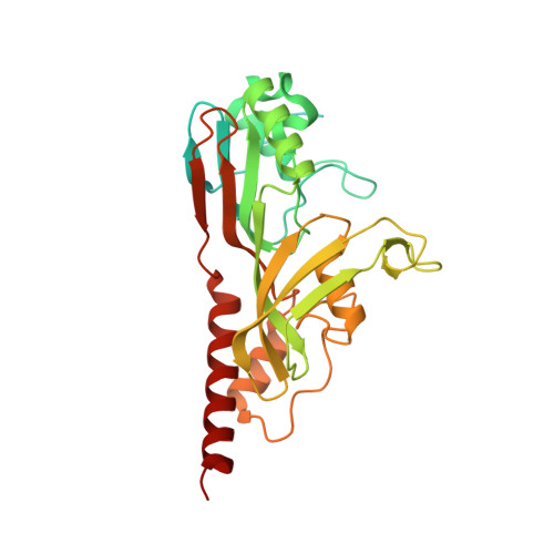
1V9F

Crystal structure of catalytic domain of pseudouridine synthase RluD from Escherichia coli


Experimental Data Snapshot

  • Method: X-RAY DIFFRACTION
  • Resolution: 1.70 Å
  • R-Value Free: 0.224 
  • R-Value Work: 0.200 
  • R-Value Observed: 0.201 

wwPDB Validation   3D Report Full Report


This is version 1.3 of the entry. See complete history


Literature

Crystal structures of the catalytic domains of pseudouridine synthases RluC and RluD from Escherichia coli

Mizutani, K.Machida, Y.Unzai, S.Park, S.-Y.Tame, J.R.H.

(2004) Biochemistry 43: 4454-4463

  • DOI: https://doi.org/10.1021/bi036079c
  • Primary Citation of Related Structures:  
    1V9F, 1V9K

  • PubMed Abstract: 

    The most frequent modification of RNA, the conversion of uridine bases to pseudouridines, is found in all living organisms and often in highly conserved locations in ribosomal and transfer RNA. RluC and RluD are homologous enzymes which each convert three specific uridine bases in Escherichia coli ribosomal 23S RNA to pseudouridine: bases 955, 2504, and 2580 in the case of RluC and 1911, 1915, and 1917 in the case of RluD. Both have an N-terminal S4 RNA binding domain. While the loss of RluC has little phenotypic effect, loss of RluD results in a much reduced growth rate. We have determined the crystal structures of the catalytic domain of RluC, and full-length RluD. The S4 domain of RluD appears to be highly flexible or unfolded and is completely invisible in the electron density map. Despite the conserved topology shared by the two proteins, the surface shape and charge distribution are very different. The models suggest significant differences in substrate binding by different pseudouridine synthases.


  • Organizational Affiliation

    Protein Design Laboratory, Yokohama City University, Suehiro 1-7-29, Tsurumi, Yokohama 230-0045, Japan.


Macromolecules
Find similar proteins by:  (by identity cutoff)  |  3D Structure
Entity ID: 1
MoleculeChains Sequence LengthOrganismDetailsImage
Ribosomal large subunit pseudouridine synthase D325Escherichia coliMutation(s): 0 
Gene Names: RLUD
EC: 4.2.1.70 (PDB Primary Data), 5.4.99.23 (UniProt)
UniProt
Find proteins for P33643 (Escherichia coli (strain K12))
Explore P33643 
Go to UniProtKB:  P33643
Entity Groups  
Sequence Clusters30% Identity50% Identity70% Identity90% Identity95% Identity100% Identity
UniProt GroupP33643
Sequence Annotations
Expand
  • Reference Sequence
Small Molecules
Ligands 1 Unique
IDChains Name / Formula / InChI Key2D Diagram3D Interactions
PO4
Query on PO4

Download Ideal Coordinates CCD File 
B [auth A]PHOSPHATE ION
O4 P
NBIIXXVUZAFLBC-UHFFFAOYSA-K
Experimental Data & Validation

Experimental Data

  • Method: X-RAY DIFFRACTION
  • Resolution: 1.70 Å
  • R-Value Free: 0.224 
  • R-Value Work: 0.200 
  • R-Value Observed: 0.201 
  • Space Group: P 65 2 2
Unit Cell:
Length ( Å )Angle ( ˚ )
a = 74.506α = 90
b = 74.506β = 90
c = 265.009γ = 120
Software Package:
Software NamePurpose
REFMACrefinement
HKL-2000data reduction
SCALEPACKdata scaling
SOLVEphasing

Structure Validation

View Full Validation Report


Entry History 

Deposition Data

Revision History  (Full details and data files)

  • Version 1.0: 2004-05-18
    Type: Initial release
  • Version 1.1: 2008-04-27
    Changes: Version format compliance
  • Version 1.2: 2011-07-13
    Changes: Version format compliance
  • Version 1.3: 2023-12-27
    Changes: Data collection, Database references, Derived calculations