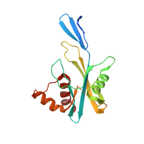
Structural insights into the Thermus thermophilus ADP-ribose pyrophosphatase mechanism via crystal structures with the bound substrate and metal
Yoshiba, S., Ooga, T., Nakagawa, N., Shibata, T., Inoue, Y., Yokoyama, S., Kuramitsu, S., Masui, R.(2004) J Biological Chem 279: 37163-37174
- PubMed: 15210687
- DOI: https://doi.org/10.1074/jbc.M403817200
- Primary Citation of Related Structures:
1V8I, 1V8L, 1V8M, 1V8N, 1V8R, 1V8S, 1V8T, 1V8U, 1V8V, 1V8W, 1V8Y - PubMed Abstract:
ADP-ribose pyrophosphatase (ADPRase) catalyzes the divalent metal ion-dependent hydrolysis of ADP-ribose to ribose 5'-phosphate and AMP. This enzyme plays a key role in regulating the intracellular ADP-ribose levels, and prevents nonenzymatic ADP-ribosylation. To elucidate the pyrophosphatase hydrolysis mechanism employed by this enzyme, structural changes occurring on binding of substrate, metal and product were investigated using crystal structures of ADPRase from an extreme thermophile, Thermus thermophilus HB8. Seven structures were determined, including that of the free enzyme, the Zn(2+)-bound enzyme, the binary complex with ADP-ribose, the ternary complexes with ADP-ribose and Zn(2+) or Gd(3+), and the product complexes with AMP and Mg(2+) or with ribose 5'-phosphate and Zn(2+). The structural and functional studies suggested that the ADP-ribose hydrolysis pathway consists of four reaction states: bound with metal (I), metal and substrate (II), metal and substrate in the transition state (III), and products (IV). In reaction state II, Glu-82 and Glu-70 abstract a proton from a water molecule. This water molecule is situated at an ideal position to carry out nucleophilic attack on the adenosyl phosphate, as it is 3.6 A away from the target phosphorus and almost in line with the scissile bond.
- Department of Biology, Graduate School of Science, Osaka University, Toyonaka, Osaka 560-0043, Japan.
Organizational Affiliation:


