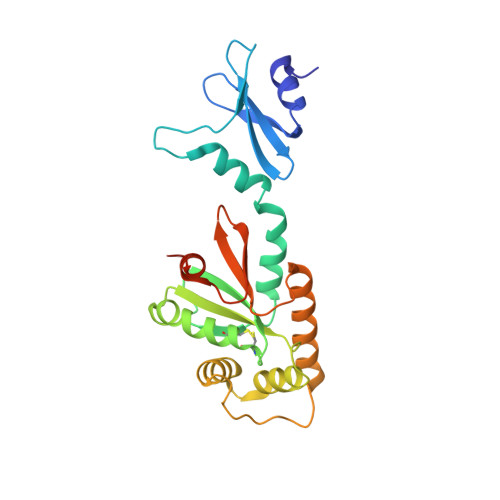
Crystal structures of the DsbG disulfide isomerase reveal an unstable disulfide
Heras, B., Edeling, M.A., Schirra, H.J., Raina, S., Martin, J.L.(2004) Proc Natl Acad Sci U S A 101: 8876-8881
- PubMed: 15184683
- DOI: https://doi.org/10.1073/pnas.0402769101
- Primary Citation of Related Structures:
1V57, 1V58 - PubMed Abstract:
Dsb proteins control the formation and rearrangement of disulfide bonds during the folding of secreted and membrane proteins in bacteria. DsbG, a member of this family, has disulfide bond isomerase and chaperone activity. Here, we present two crystal structures of DsbG at 1.7and 2.0-A resolution that are meant to represent the reduced and oxidized forms, respectively. The oxidized structure, however, reveals a mixture of both redox forms, suggesting that oxidized DsbG is less stable than the reduced form. This trait would contribute to DsbG isomerase activity, which requires that the active-site Cys residues are kept reduced, regardless of the highly oxidative environment of the periplasm. We propose that a Thr residue that is conserved in the cis-Pro loop of DsbG and DsbC but not found in other Dsb proteins could play a role in this process. Also, the structure of DsbG reveals an unanticipated and surprising feature that may help define its specific role in oxidative protein folding. Thus, the dimensions and surface features of DsbG show a very large and charged binding surface that is consistent with interaction with globular protein substrates having charged surfaces. This finding suggests that, rather than catalyzing disulfide rearrangement in unfolded substrates, DsbG may preferentially act later in the folding process to catalyze disulfide rearrangement in folded or partially folded proteins.
- Institute for Molecular Bioscience and Australian Research Council Special Research Centre for Functional and Applied Genomics, University of Queensland, Brisbane QLD 4072, Australia.
Organizational Affiliation:


