1V3W

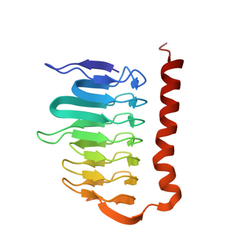
Structure of Ferripyochelin binding protein from Pyrococcus horikoshii OT3


Experimental Data Snapshot

  • Method: X-RAY DIFFRACTION
  • Resolution: 1.50 Å
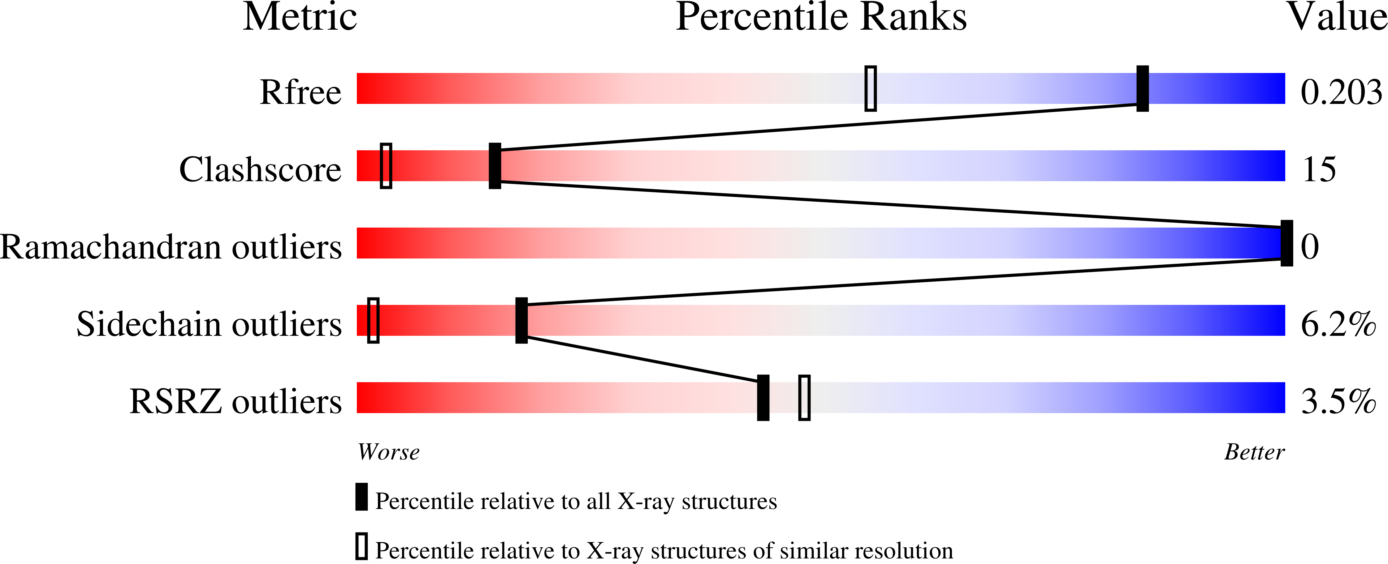
  • R-Value Free: 0.203 
  • R-Value Work: 0.185 

Starting Model: experimental
View more details

wwPDB Validation   3D Report Full Report


This is version 1.4 of the entry. See complete history


Literature

Observation of a calcium-binding site in the gamma-class carbonic anhydrase from Pyrococcus horikoshii.

Jeyakanthan, J.Rangarajan, S.Mridula, P.Kanaujia, S.P.Shiro, Y.Kuramitsu, S.Yokoyama, S.Sekar, K.

(2008) Acta Crystallogr D Biol Crystallogr 64: 1012-1019

  • DOI: https://doi.org/10.1107/S0907444908024323
  • Primary Citation of Related Structures:  
    1V3W, 1V67, 2FKO

  • PubMed Abstract: 

    Carbonic anhydrases are zinc-containing metalloenzymes that catalyze the interconversion of carbon dioxide and bicarbonate. Three crystal structures of gamma-class carbonic anhydrase (one of which is bound to a bicarbonate molecule) from the aerobic OT3 strain of the hyperthermophilic archeon Pyrococcus horikoshii have been solved by molecular replacement in space group F4(1)32. The asymmetric unit contains a monomer of 173 amino acids and a catalytic Zn2+ ion. The protein fold is a regular prism formed by a left-handed beta-helix, similar to previously reported structures. The active-site Zn2+ ion located at the interface between the two monomers is bound to three histidyl residues and a water molecule in a tetrahedral fashion. In addition to the 20 beta-strands comprising the beta-helix, there is also a long C-terminal alpha-helix. For the first time, Ca2+ ions have been observed in addition to the catalytic Zn2+ ion. It is hypothesized that Tyr159 (which corresponds to the catalytically important Asn202 in previously reported structures) utilizes C-H...pi interactions to fulfill its functions. This study may shed light on the catalytic mechanism of the enzyme and throw open new questions on the mechanism of product removal in carbonic anhydrases.


  • Organizational Affiliation

    National Synchrotron Radiation Research Center, 101 Hsin-Ann Road, Hsinchu Science Park, Hsinchu 30076, Taiwan.


Macromolecules
Find similar proteins by:  (by identity cutoff)  |  3D Structure
Entity ID: 1
MoleculeChains Sequence LengthOrganismDetailsImage
ferripyochelin binding protein173Pyrococcus horikoshii OT3Mutation(s): 0 
EC: 4.2.1.1
UniProt
Find proteins for O59257 (Pyrococcus horikoshii (strain ATCC 700860 / DSM 12428 / JCM 9974 / NBRC 100139 / OT-3))
Explore O59257 
Go to UniProtKB:  O59257
Entity Groups  
Sequence Clusters30% Identity50% Identity70% Identity90% Identity95% Identity100% Identity
UniProt GroupO59257
Sequence Annotations
Expand
  • Reference Sequence
Small Molecules
Ligands 5 Unique
IDChains Name / Formula / InChI Key2D Diagram3D Interactions
GOL
Query on GOL

Download Ideal Coordinates CCD File 
K [auth A]GLYCEROL
C3 H8 O3
PEDCQBHIVMGVHV-UHFFFAOYSA-N
ZN
Query on ZN

Download Ideal Coordinates CCD File 
B [auth A]ZINC ION
Zn
PTFCDOFLOPIGGS-UHFFFAOYSA-N
EDO
Query on EDO

Download Ideal Coordinates CCD File 
H [auth A],
I [auth A],
J [auth A]
1,2-ETHANEDIOL
C2 H6 O2
LYCAIKOWRPUZTN-UHFFFAOYSA-N
CA
Query on CA

Download Ideal Coordinates CCD File 
C [auth A],
D [auth A],
E [auth A],
F [auth A]
CALCIUM ION
Ca
BHPQYMZQTOCNFJ-UHFFFAOYSA-N
CL
Query on CL

Download Ideal Coordinates CCD File 
G [auth A]CHLORIDE ION
Cl
VEXZGXHMUGYJMC-UHFFFAOYSA-M
Experimental Data & Validation

Experimental Data

  • Method: X-RAY DIFFRACTION
  • Resolution: 1.50 Å
  • R-Value Free: 0.203 
  • R-Value Work: 0.185 
  • Space Group: F 41 3 2
Unit Cell:
Length ( Å )Angle ( ˚ )
a = 156.156α = 90
b = 156.156β = 90
c = 156.156γ = 90
Software Package:
Software NamePurpose
HKL-2000data collection
SCALEPACKdata scaling
AMoREphasing
CNSrefinement
HKL-2000data reduction

Structure Validation

View Full Validation Report


Entry History 

Deposition Data

Revision History  (Full details and data files)

  • Version 1.0: 2003-11-18
    Type: Initial release
  • Version 1.1: 2008-04-27
    Changes: Version format compliance
  • Version 1.2: 2011-07-13
    Changes: Derived calculations, Source and taxonomy, Version format compliance
  • Version 1.3: 2013-01-30
    Changes: Structure summary
  • Version 1.4: 2023-10-25
    Changes: Data collection, Database references, Derived calculations, Refinement description