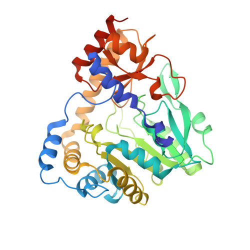
1V2E

Crystal Structure of T.th HB8 Glutamine Aminotransferase complex with a-keto-g-methylthiobutyrate


Experimental Data Snapshot

  • Method: X-RAY DIFFRACTION
  • Resolution: 2.60 Å
  • R-Value Free: 0.286 
  • R-Value Work: 0.231 

wwPDB Validation   3D Report Full Report


Ligand Structure Quality Assessment 

Created with Raphaël 2.3.0Worse 01 BetterLigand structure goodness of fit to experimental dataBest fitted PLPClick on this verticalbar to view details

This is version 1.3 of the entry. See complete history


Literature

Crystal structures of glutamine:phenylpyruvate aminotransferase from Thermus thermophilus HB8: induced fit and substrate recognition

Goto, M.Omi, R.Miyahara, I.Hosono, A.Mizuguchi, H.Hayashi, H.Kagamiyama, H.Hirotsu, K.

(2004) J Biol Chem 279: 16518-16525

  • DOI: https://doi.org/10.1074/jbc.M311575200
  • Primary Citation of Related Structures:  
    1V2D, 1V2E, 1V2F

  • PubMed Abstract: 

    The following three-dimensional structures of three forms of glutamine:phenylpyruvate aminotransferase from Thermus thermophilus HB8 have been determined and represent the first x-ray analysis of the enzyme: the unliganded pyridoxal 5'-phosphate form at 1.9 A resolution and two complexes with 3-phenylpropionate and alpha-keto-gamma-methylthiobutyrate at 2.35 and 2.6 A resolution, respectively. The enzyme shows high activity toward phenylalanine, tyrosine, tryptophan, kynurenine, methionine, and glutamine. The enzyme is a homodimer, and each subunit is divided into an N-terminal arm and small and large domains. Based on its folding, the enzyme belongs to fold type I, aminotransferase subclass Ib. The subclass I aminotransferases whose structures have so far been determined exhibit a large movement of the small domain region upon binding of a substrate. Similarly, the glutamine:phenylpyruvate aminotransferase undergoes a large movement in part of the small domain to close the active site. The active-site pocket has a shape and size suitable to enclose the side chain of an aromatic amino acid or that of methionine. The inner side of the pocket is mostly hydrophobic, but also has polar sites. The kynurenine complex generated by computer modeling fits the pocket of the enzyme and its hydrophilic groups interact with the polar sites of the pocket.


  • Organizational Affiliation

    Department of Chemistry, Graduate School of Science, Osaka City University, Osaka 558-8585, Japan.


Macromolecules
Find similar proteins by:  (by identity cutoff)  |  3D Structure
Entity ID: 1
MoleculeChains Sequence LengthOrganismDetailsImage
Glutamine Aminotransferase
A, B
381Thermus thermophilusMutation(s): 0 
Gene Names: HB8
EC: 2.6.1.15
UniProt
Find proteins for Q75WK2 (Thermus thermophilus)
Explore Q75WK2 
Go to UniProtKB:  Q75WK2
Entity Groups  
Sequence Clusters30% Identity50% Identity70% Identity90% Identity95% Identity100% Identity
UniProt GroupQ75WK2
Sequence Annotations
Expand
  • Reference Sequence
Experimental Data & Validation

Experimental Data

  • Method: X-RAY DIFFRACTION
  • Resolution: 2.60 Å
  • R-Value Free: 0.286 
  • R-Value Work: 0.231 
  • Space Group: P 31 2 1
Unit Cell:
Length ( Å )Angle ( ˚ )
a = 123.14α = 90
b = 123.14β = 90
c = 122.96γ = 120
Software Package:
Software NamePurpose
MOSFLMdata reduction
SCALAdata scaling
AMoREphasing
CNSrefinement
CCP4data scaling

Structure Validation

View Full Validation Report


Ligand Structure Quality Assessment 

Created with Raphaël 2.3.0Worse 01 BetterLigand structure goodness of fit to experimental dataBest fitted PLPClick on this verticalbar to view details

Entry History 

Deposition Data

Revision History  (Full details and data files)

  • Version 1.0: 2004-07-06
    Type: Initial release
  • Version 1.1: 2008-04-27
    Changes: Version format compliance
  • Version 1.2: 2011-07-13
    Changes: Non-polymer description, Version format compliance
  • Version 1.3: 2023-12-27
    Changes: Data collection, Database references, Derived calculations