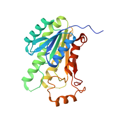
Crystal Structure of Maba from Mycobacterium Tuberculosis, a Reductase Involved in Long-Chain Fatty Acid Biosynthesis.
Cohen-Gonsaud, M., Ducasse, S., Hoh, F., Zerbib, D., Labesse, G., Quemard, A.(2002) J Mol Biology 320: 249
- PubMed: 12079383
- DOI: https://doi.org/10.1016/S0022-2836(02)00463-1
- Primary Citation of Related Structures:
1UZL, 1UZM, 1UZN - PubMed Abstract:
The fatty acid elongation system FAS-II is involved in the biosynthesis of mycolic acids, which are major and specific long-chain fatty acids of the cell envelope of Mycobacterium tuberculosis and other mycobacteria, including Mycobacterium smegmatis. The protein MabA, also named FabG1, has been shown recently to be part of FAS-II and to catalyse the NADPH-specific reduction of long chain beta-ketoacyl derivatives. This activity corresponds to the second step of an FAS-II elongation round. FAS-II is inhibited by the antituberculous drug isoniazid through the inhibition of the 2-trans-enoyl-acyl carrier protein reductase InhA. Thus, the other enzymes making up this enzymatic complex represent potential targets for designing new antituberculous drugs. The crystal structure of the apo-form MabA was solved to 2.03 A resolution by molecular replacement. MabA is tetrameric and shares the conserved fold of the short-chain dehydrogenases/reductases (SDRs). However, it exhibits some significant local rearrangements of the active-site loops in the absence of a cofactor, particularly the beta5-alpha5 region carrying the unique tryptophan residue, in agreement with previous fluorescence spectroscopy data. A similar conformation has been observed in the beta-ketoacyl reductase from Escherichia coli and the distantly related dehydratase. The distinctive enzymatic and structural properties of MabA are discussed in view of its crystal structure and that of related enzymes.
- Centre de Biochimie Structurale (INSERM U554-CNRS UMR5048-UM1), 29 rue de Navacelles, 34090 Montpellier cedex, France. martin@cbs.cnrs.fr
Organizational Affiliation:


