1UZA | pdb_00001uza

Crystallographic structure of a feruloyl esterase from Aspergillus niger


Experimental Data Snapshot

  • Method: X-RAY DIFFRACTION
  • Resolution: 1.50 Å
  • R-Value Free: 
    0.188 (Depositor), 0.195 (DCC) 
  • R-Value Work: 
    0.156 (Depositor), 0.166 (DCC) 
  • R-Value Observed: 
    0.158 (Depositor) 

Starting Model: experimental
View more details

wwPDB Validation 3D Report Full Report

Validation slider image for 1UZA

Ligand Structure Quality Assessment 


This is version 1.4 of the entry. See complete history

Literature

Structure of a Feruloyl Esterase from Aspergillus Niger

Mcauley, K.E.Svendsen, A.Patkar, S.A.Wilson, K.S.

(2004) Acta Crystallogr D Biol Crystallogr 60: 878

  • DOI: https://doi.org/10.1107/S0907444904004937
  • Primary Citation Related Structures: 
    1UWC, 1UZA

  • PubMed Abstract: 

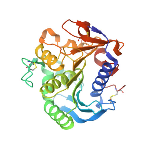
    The crystallographic structure of feruloyl esterase from Aspergillus niger has been determined to a resolution of 1.5 A by molecular replacement. The protein has an alpha/beta-hydrolase structure with a Ser-His-Asp catalytic triad; the overall fold of the protein is very similar to that of the fungal lipases. The structure of the enzyme-product complex was determined to a resolution of 1.08 A and reveals dual conformations for the serine and histidine residues at the active site.


  • Organizational Affiliation
    • CCLRC Daresbury Laboratory, Warrington, Cheshire WA4 4AD, England.

Macromolecule Content 

  • Total Structure Weight: 57.58 kDa 
  • Atom Count: 4,475 
  • Modeled Residue Count: 520 
  • Deposited Residue Count: 522 
  • Unique protein chains: 1

Macromolecules

Find similar proteins by:|  3D Structure
Entity ID: 1
MoleculeChains  Sequence LengthOrganismDetailsImage
FERULOYL ESTERASE A
A, B
261Aspergillus nigerMutation(s): 0 
EC: 3.1.1.73
UniProt
Find proteins for O42807 (Aspergillus niger)
Explore O42807 
Go to UniProtKB:  O42807
Entity Groups
Sequence Clusters30% Identity50% Identity70% Identity90% Identity95% Identity100% Identity
UniProt GroupO42807
Glycosylation
Glycosylation Sites: 1
Sequence Annotations
Expand
Reference Sequence

Experimental Data & Validation

Experimental Data

  • Method: X-RAY DIFFRACTION
  • Resolution: 1.50 Å
  • R-Value Free:  0.188 (Depositor), 0.195 (DCC) 
  • R-Value Work:  0.156 (Depositor), 0.166 (DCC) 
  • R-Value Observed: 0.158 (Depositor) 
Space Group: P 1
Unit Cell:
Length ( Å )Angle ( ˚ )
a = 38.298α = 75.24
b = 39.681β = 78.82
c = 77.071γ = 71.3
Software Package:
Software NamePurpose
REFMACrefinement
DENZOdata reduction
SCALEPACKdata scaling
AMoREphasing

Structure Validation

View Full Validation Report


Ligand Structure Quality Assessment 


Entry History 

Deposition Data

Revision History  (Full details and data files)

  • Version 1.0: 2004-10-19
    Type: Initial release
  • Version 1.1: 2012-11-14
    Changes: Database references, Derived calculations, Non-polymer description, Other, Version format compliance
  • Version 1.2: 2020-07-29
    Type: Remediation
    Reason: Carbohydrate remediation
    Changes: Data collection, Derived calculations, Other, Structure summary
  • Version 1.3: 2023-12-13
    Changes: Data collection, Database references, Refinement description, Structure summary
  • Version 1.4: 2024-11-13
    Changes: Structure summary