1UZ8 | pdb_00001uz8

anti-Lewis X Fab fragment in complex with Lewis X


Experimental Data Snapshot

  • Method: X-RAY DIFFRACTION
  • Resolution: 1.80 Å
  • R-Value Free: 
    0.247 (Depositor), 0.288 (DCC) 
  • R-Value Work: 
    0.210 (Depositor), 0.264 (DCC) 

Starting Model: experimental
View more details

wwPDB Validation 3D Report Full Report

Validation slider image for 1UZ8

This is version 2.2 of the entry. See complete history

Literature

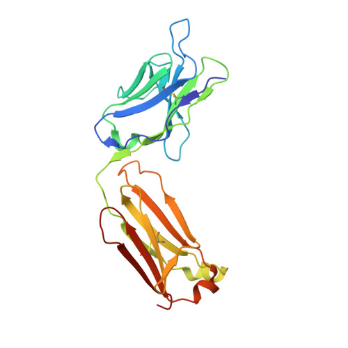
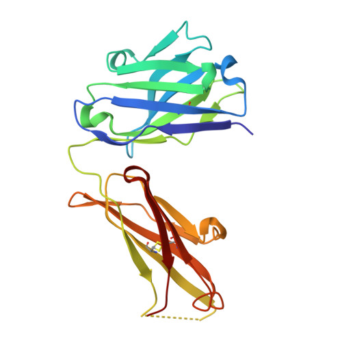
Structure of an Anti-Lewis X Fab Fragment in Complex with its Lewis X Antigen

Van Roon, A.M.M.Pannu, N.S.De Vrind, J.P.M.Van Der Marel, G.A.Van Boom, J.H.Hokke, C.H.Deelder, A.M.Abrahams, J.P.

(2004) Structure 12: 1227

  • DOI: https://doi.org/10.1016/j.str.2004.05.008
  • Primary Citation Related Structures: 
    1UZ6, 1UZ8

  • PubMed Abstract: 

    The Lewis X trisaccharide is pivotal in mediating specific cell-cell interactions. Monoclonal antibody 291-2G3-A, which was generated from mice infected with schistosomes, has been shown to recognize the Lewis X trisaccharide. Here we describe the structure of the Fab fragment of 291-2G3-A, with Lewis X, to 1.8 A resolution. The crystallographic analysis revealed that the antigen binding site is a rather shallow binding pocket, and residues from all six complementary determining regions of the antibody contact all sugar residues. The high specificity of the binding pocket does not result in high affinity; the K(D) determined by isothermal calorimetry is 11 microM. However, this affinity is in the same range as for other sugar-antibody complexes. The detailed understanding of the antibody-Lewis X interaction revealed by the crystal structure may be helpful in the design of better diagnostic tools for schistosomiasis and for studying Lewis X-mediated cell-cell interactions by antibody interference.


  • Organizational Affiliation
    • Department of Biophysical Structural Chemistry, Leiden University, P.O. Box 9502, 2300 RA Leiden, The Netherlands.

Macromolecule Content 

  • Total Structure Weight: 95.09 kDa 
  • Atom Count: 7,002 
  • Modeled Residue Count: 860 
  • Deposited Residue Count: 860 
  • Unique protein chains: 2

Macromolecules

Find similar proteins by:|  3D Structure
Entity ID: 1
MoleculeChains  Sequence LengthOrganismDetailsImage
IGG FAB (IGG3, KAPPA) LIGHT CHAIN 291-2G3-AA,
D [auth L]
218Mus musculusMutation(s): 0 
Find similar proteins by:|  3D Structure
Entity ID: 2
MoleculeChains  Sequence LengthOrganismDetailsImage
IGG FAB (IGG3, KAPPA) HEAVY CHAIN 291-2G3-AB,
C [auth H]
212Mus musculusMutation(s): 0 
UniProt
Find proteins for P01811 (Mus musculus)
Explore P01811 
Go to UniProtKB:  P01811
Entity Groups
Sequence Clusters30% Identity50% Identity70% Identity90% Identity95% Identity100% Identity
UniProt GroupP01811
Sequence Annotations
Expand
Reference Sequence

Oligosaccharides

Help  
Entity ID: 3
MoleculeChains Length2D Diagram GlycosylationD Interactions
alpha-L-fucopyranose-(1-3)-[beta-D-galactopyranose-(1-4)]methyl 2-acetamido-2-deoxy-beta-D-glucopyranosideE [auth C],
F [auth D]
3N/A
Glycosylation Resources
GlyTouCan: G26481RZ
GlyCosmos: G26481RZ
GlyGen: G26481RZ

Experimental Data & Validation

Experimental Data

  • Method: X-RAY DIFFRACTION
  • Resolution: 1.80 Å
  • R-Value Free:  0.247 (Depositor), 0.288 (DCC) 
  • R-Value Work:  0.210 (Depositor), 0.264 (DCC) 
Space Group: P 1
Unit Cell:
Length ( Å )Angle ( ˚ )
a = 45.104α = 95.98
b = 60.778β = 95.41
c = 91.556γ = 101.71
Software Package:
Software NamePurpose
REFMACrefinement
HKL-2000data reduction
SCALEPACKdata scaling
MOLREPphasing

Structure Validation

View Full Validation Report


Entry History 

Deposition Data

Revision History  (Full details and data files)

  • Version 1.0: 2004-06-29
    Type: Initial release
  • Version 1.1: 2011-07-13
    Changes: Advisory, Version format compliance
  • Version 2.0: 2020-07-29
    Type: Remediation
    Reason: Carbohydrate remediation
    Changes: Atomic model, Data collection, Derived calculations, Other, Structure summary
  • Version 2.1: 2023-12-13
    Changes: Data collection, Database references, Refinement description, Structure summary
  • Version 2.2: 2024-10-16
    Changes: Structure summary