1UV5 | pdb_00001uv5

GLYCOGEN SYNTHASE KINASE 3 BETA COMPLEXED WITH 6-BROMOINDIRUBIN-3'-OXIME


Experimental Data Snapshot

  • Method: X-RAY DIFFRACTION
  • Resolution: 2.80 Å
  • R-Value Free: 
    0.226 (Depositor) 
  • R-Value Work: 
    0.193 (Depositor) 
  • R-Value Observed: 
    0.193 (Depositor) 

Starting Model: experimental
View more details

wwPDB Validation 3D Report Full Report

Validation slider image for 1UV5

Ligand Structure Quality Assessment 


This is version 1.1 of the entry. See complete history

Literature

Gsk-3-Selective Inhibitors Derived from Tyrian Purple Indurubins

Meijer, L.Skaltsounis, A.-L.Magiatis, P.Polychronopoulous, P.Knockaert, M.Leost, M.Ryan, X.P.Vonica, C.A.Brivanlou, A.Dajani, R.Crovace, C.Tarricone, C.Musacchio, A.Roe, S.M.Pearl, L.H.Greengard, P.

(2003) Chem Biol 10: 1255

  • DOI: https://doi.org/10.1016/j.chembiol.2003.11.010
  • Primary Citation Related Structures: 
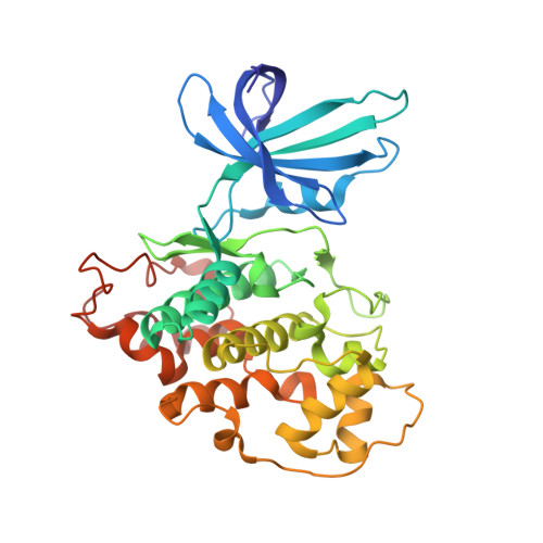
    1UV5

  • PubMed Abstract: 

    Gastropod mollusks have been used for over 2500 years to produce the "Tyrian purple" dye made famous by the Phoenicians. This dye is constituted of mixed bromine-substituted indigo and indirubin isomers. Among these, the new natural product 6-bromoindirubin and its synthetic, cell-permeable derivative, 6-bromoindirubin-3'-oxime (BIO), display remarkable selective inhibition of glycogen synthase kinase-3 (GSK-3). Cocrystal structure of GSK-3beta/BIO and CDK5/p25/indirubin-3'-oxime were resolved, providing a detailed view of indirubins' interactions within the ATP binding pocket of these kinases. BIO but not 1-methyl-BIO, its kinase inactive analog, also inhibited the phosphorylation on Tyr276/216, a GSK-3alpha/beta activation site. BIO but not 1-methyl-BIO reduced beta-catenin phosphorylation on a GSK-3-specific site in cellular models. BIO but not 1-methyl-BIO closely mimicked Wnt signaling in Xenopus embryos. 6-bromoindirubins thus provide a new scaffold for the development of selective and potent pharmacological inhibitors of GSK-3.


  • Organizational Affiliation
    • CNRS, Cell Cycle Group, Station Biologique, BP 74, 29682 Roscoff cedex, Bretagne, France. meijer@sb-roscoff.fr

Macromolecule Content 

  • Total Structure Weight: 40.3 kDa 
  • Atom Count: 2,906 
  • Modeled Residue Count: 349 
  • Deposited Residue Count: 350 
  • Unique protein chains: 1

Macromolecules

Find similar proteins by:|  3D Structure
Entity ID: 1
MoleculeChains  Sequence LengthOrganismDetailsImage
GLYCOGEN SYNTHASE KINASE-3 BETA350Homo sapiensMutation(s): 0 
EC: 2.7.1.37 (PDB Primary Data), 2.7.11.1 (UniProt), 2.7.11.26 (UniProt)
UniProt & NIH Common Fund Data Resources
Find proteins for P49841 (Homo sapiens)
Explore P49841 
Go to UniProtKB:  P49841
PHAROS:  P49841
GTEx:  ENSG00000082701 
Entity Groups
Sequence Clusters30% Identity50% Identity70% Identity90% Identity95% Identity100% Identity
UniProt GroupP49841
Sequence Annotations
Expand
Reference Sequence

Small Molecules

Binding Affinity Annotations 
IDSourceBinding Affinity
CL BindingDB:  1UV5 IC50: 6530 (nM) from 1 assay(s)
EC50: 3.00e+6 (nM) from 1 assay(s)
BRW BindingDB:  1UV5 IC50: 5 (nM) from 1 assay(s)

Experimental Data & Validation

Experimental Data

  • Method: X-RAY DIFFRACTION
  • Resolution: 2.80 Å
  • R-Value Free:  0.226 (Depositor) 
  • R-Value Work:  0.193 (Depositor) 
  • R-Value Observed: 0.193 (Depositor) 
Space Group: P 43 21 2
Unit Cell:
Length ( Å )Angle ( ˚ )
a = 98.333α = 90
b = 98.333β = 90
c = 198.017γ = 90
Software Package:
Software NamePurpose
CNSrefinement
MOSFLMdata reduction
SCALAdata scaling
AMoREphasing

Structure Validation

View Full Validation Report


Ligand Structure Quality Assessment 


Entry History 

Deposition Data

Revision History  (Full details and data files)

  • Version 1.0: 2004-01-29
    Type: Initial release
  • Version 1.1: 2023-12-13
    Changes: Data collection, Database references, Derived calculations, Other, Refinement description