1UIJ | pdb_00001uij

Crystal Structure Of Soybean beta-Conglycinin Beta Homotrimer (I122M/K124W)


Experimental Data Snapshot

  • Method: X-RAY DIFFRACTION
  • Resolution: 2.50 Å
  • R-Value Free: 
    0.273 (Depositor), 0.276 (DCC) 
  • R-Value Work: 
    0.221 (Depositor), 0.225 (DCC) 

wwPDB Validation 3D Report Full Report

Validation slider image for 1UIJ

This is version 1.4 of the entry. See complete history

Literature

Creation of soybean beta-conglycinin beta with strong phagocytosis-stimulating activity

Maruyama, N.Maruyama, Y.Tsuruki, T.Okuda, E.Yoshikawa, M.Utsumi, S.

(2003) Biochim Biophys Acta 1648: 99-104

  • DOI: https://doi.org/10.1016/s1570-9639(03)00113-4
  • Primary Citation Related Structures: 
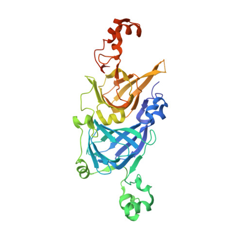
    1UIJ

  • PubMed Abstract: 

    beta-Conglycinin is composed of three kinds of subunit: alpha, alpha' and beta. A phagocytosis-stimulating peptide sequence (MITLAIPVNKPGR), soymetide, exists in the alpha' subunit of beta-conglycinin. Met at N terminus of the soymetide is essential for the activity. When Thr at the third residue from N terminus of the soymetide is replaced by Phe or Trp, the phagocytosis-stimulating activity greatly increases (ThrMet, Lys-->Thr, Phe, or Trp) into the beta subunit after confirmation of the effects of residue replacements by molecular modeling, suggesting that the introduced mutations might not prevent the correct folding. The studies of circular dichroism (CD), gel filtration and differential scanning calorimetry (DSC) of the mutants (I122M/K124T, I122M/K124F, I122M/K124W) expressed in E. coli demonstrated that they folded and self-assembled similarly to the wild type. This was confirmed by X-ray analysis of I122M/K124W crystal where the biggest residue tryptophane was introduced. The three mutants exhibited phagocytosis activities after digestion by trypsin, and the order was the wild type


  • Organizational Affiliation
    • Laboratory of Food Quality Design and Development, Graduate School of Agriculture, Kyoto University, Uji, Japan.

Macromolecule Content 

  • Total Structure Weight: 288.65 kDa 
  • Atom Count: 18,133 
  • Modeled Residue Count: 2,186 
  • Deposited Residue Count: 2,496 
  • Unique protein chains: 1

Macromolecules

Find similar proteins by:|  3D Structure
Entity ID: 1
MoleculeChains  Sequence LengthOrganismDetailsImage
beta subunit of beta conglycinin
A, B, C, D, E
A, B, C, D, E, F
416Glycine maxMutation(s): 2 
UniProt
Find proteins for P25974 (Glycine max)
Explore P25974 
Go to UniProtKB:  P25974
Entity Groups
Sequence Clusters30% Identity50% Identity70% Identity90% Identity95% Identity100% Identity
UniProt GroupP25974
Sequence Annotations
Expand
Reference Sequence

Experimental Data & Validation

Experimental Data

  • Method: X-RAY DIFFRACTION
  • Resolution: 2.50 Å
  • R-Value Free:  0.273 (Depositor), 0.276 (DCC) 
  • R-Value Work:  0.221 (Depositor), 0.225 (DCC) 
Space Group: P 1 21 1
Unit Cell:
Length ( Å )Angle ( ˚ )
a = 130.263α = 90
b = 62.583β = 90.44
c = 159.009γ = 90
Software Package:
Software NamePurpose
HKL-2000data collection
SCALEPACKdata scaling
AMoREphasing
CNSrefinement
HKL-2000data reduction

Structure Validation

View Full Validation Report


Entry History 

Deposition Data

Revision History  (Full details and data files)

  • Version 1.0: 2004-07-16
    Type: Initial release
  • Version 1.1: 2008-04-27
    Changes: Version format compliance
  • Version 1.2: 2011-07-13
    Changes: Version format compliance
  • Version 1.3: 2021-11-10
    Changes: Database references
  • Version 1.4: 2023-12-27
    Changes: Data collection