1UFI | pdb_00001ufi

Crystal structure of the dimerization domain of human CENP-B


Experimental Data Snapshot

  • Method: X-RAY DIFFRACTION
  • Resolution: 1.65 Å
  • R-Value Free: 
    0.309 (Depositor), 0.232 (DCC) 
  • R-Value Work: 
    0.232 (Depositor), 0.240 (DCC) 
  • R-Value Observed: 
    0.236 (Depositor) 

wwPDB Validation 3D Report Full Report

Validation slider image for 1UFI

This is version 1.3 of the entry. See complete history

Literature

Crystal structure of the human centromere protein B (CENP-B) dimerization domain at 1.65-A resolution

Tawaramoto, M.S.Park, S.-Y.Tanaka, Y.Nureki, O.Kurumizaka, H.Yokoyama, S.

(2003) J Biological Chem 278: 51454-51461

  • DOI: https://doi.org/10.1074/jbc.M310388200
  • Primary Citation Related Structures: 
    1UFI

  • PubMed Abstract: 

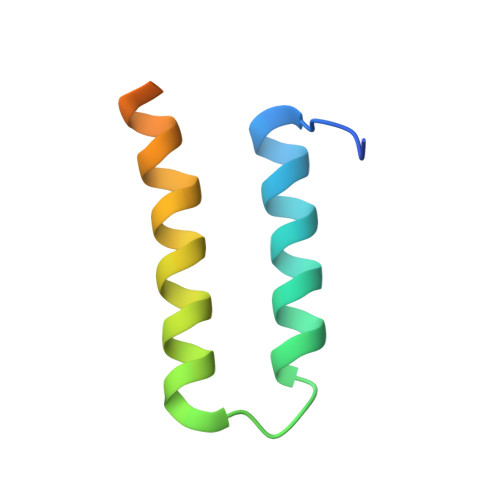
    The human centromere protein B (CENP-B), a centromeric heterochromatin component, forms a homodimer that specifically binds to a distinct DNA sequence (the CENP-B box), which appears within every other alpha-satellite repeat. Previously, we determined the structure of the human CENP-B DNA-binding domain, CENP-B-(1-129), complexed with the CENP-B box DNA. In the present study, we determined the crystal structure of its dimerization domain (CENP-B-(540-599)), another functional domain of CENP-B, at 1.65-A resolution. CENP-B-(540-599) contains two alpha-helices, which are folded into an antiparallel configuration. The CENP-B-(540-599) dimer formed a symmetrical, antiparallel, four-helix bundle structure with a large hydrophobic patch in which 23 residues of one monomer form van der Waals contacts with the other monomer. In the CENP-B-(540-599) dimer, the N-terminal ends of CENP-B-(540-599) are oriented on opposite sides of the dimer. This CENP-B dimer configuration may be suitable for capturing two distant CENP-B boxes during centromeric heterochromatin formation.


  • Organizational Affiliation
    • RIKEN Genomic Sciences Center, 1-7-22 Suehiro-cho, Tsurumi, Yokohama 230-0045, Japan.

Macromolecule Content 

  • Total Structure Weight: 29 kDa 
  • Atom Count: 1,694 
  • Modeled Residue Count: 188 
  • Deposited Residue Count: 256 
  • Unique protein chains: 1

Macromolecules

Find similar proteins by:|  3D Structure
Entity ID: 1
MoleculeChains  Sequence LengthOrganismDetailsImage
Major centromere autoantigen B
A, B, C, D
64Homo sapiensMutation(s): 0 
Gene Names: CENPB
UniProt & NIH Common Fund Data Resources
Find proteins for P07199 (Homo sapiens)
Explore P07199 
Go to UniProtKB:  P07199
PHAROS:  P07199
GTEx:  ENSG00000125817 
Entity Groups
Sequence Clusters30% Identity50% Identity70% Identity90% Identity95% Identity100% Identity
UniProt GroupP07199
Sequence Annotations
Expand
Reference Sequence

Experimental Data & Validation

Experimental Data

  • Method: X-RAY DIFFRACTION
  • Resolution: 1.65 Å
  • R-Value Free:  0.309 (Depositor), 0.232 (DCC) 
  • R-Value Work:  0.232 (Depositor), 0.240 (DCC) 
  • R-Value Observed: 0.236 (Depositor) 
Space Group: P 21 21 21
Unit Cell:
Length ( Å )Angle ( ˚ )
a = 43.707α = 90
b = 48.955β = 90
c = 100.7γ = 90
Software Package:
Software NamePurpose
REFMACrefinement
HKL-2000data reduction
SCALEPACKdata scaling
SOLVEphasing

Structure Validation

View Full Validation Report


Entry History 

Revision History  (Full details and data files)

  • Version 1.0: 2004-02-17
    Type: Initial release
  • Version 1.1: 2008-04-27
    Changes: Version format compliance
  • Version 1.2: 2011-07-13
    Changes: Version format compliance
  • Version 1.3: 2023-12-27
    Changes: Data collection, Database references