1UAL | pdb_00001ual

Crystal structure of tRNA(m1G37)methyltransferase: Insight into tRNA recognition


Experimental Data Snapshot

  • Method: X-RAY DIFFRACTION
  • Resolution: 1.80 Å
  • R-Value Free: 
    0.215 (Depositor), 0.215 (DCC) 
  • R-Value Work: 
    0.184 (Depositor), 0.184 (DCC) 
  • R-Value Observed: 
    0.184 (Depositor) 

wwPDB Validation 3D Report Full Report

Validation slider image for 1UAL

Ligand Structure Quality Assessment 


This is version 1.3 of the entry. See complete history

Literature

Crystal structure of tRNA(m(1)G37)methyltransferase: insights into tRNA recognition

Ahn, H.J.Kim, H.-W.Yoon, H.-J.Lee, B.I.Suh, S.W.Yang, J.K.

(2003) EMBO J 22: 2593-2603

  • DOI: https://doi.org/10.1093/emboj/cdg269
  • Primary Citation Related Structures: 
    1UAJ, 1UAK, 1UAL, 1UAM

  • PubMed Abstract: 

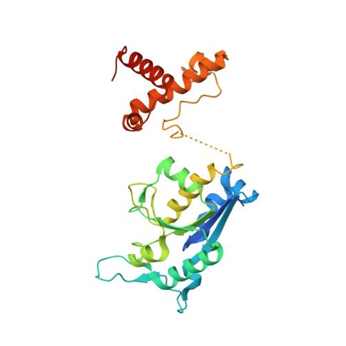
    tRNA(m(1)G37)methyltransferase (TrmD) catalyzes the transfer of a methyl group from S-adenosyl-L- methionine (AdoMet) to G(37) within a subset of bacterial tRNA species, which have a G residue at the 36th position. The modified guanosine is adjacent to and 3' of the anticodon and is essential for the maintenance of the correct reading frame during translation. Here we report four crystal structures of TrmD from Haemophilus influenzae, as binary complexes with either AdoMet or S-adenosyl-L-homocysteine (AdoHcy), as a ternary complex with AdoHcy and phosphate, and as an apo form. This first structure of TrmD indicates that it functions as a dimer. It also suggests the binding mode of G(36)G(37) in the active site of TrmD and the catalytic mechanism. The N-terminal domain has a trefoil knot, in which AdoMet or AdoHcy is bound in a novel, bent conformation. The C-terminal domain shows structural similarity to trp repressor. We propose a plausible model for the TrmD(2)-tRNA(2) complex, which provides insights into recognition of the general tRNA structure by TrmD.


  • Organizational Affiliation
    • Structural Proteomics Laboratory, School of Chemistry and Molecular Engineering, Seoul National University, Seoul 151-742, Korea.

Macromolecule Content 

  • Total Structure Weight: 31.21 kDa 
  • Atom Count: 2,229 
  • Modeled Residue Count: 243 
  • Deposited Residue Count: 274 
  • Unique protein chains: 1

Macromolecules

Find similar proteins by:|  3D Structure
Entity ID: 1
MoleculeChains  Sequence LengthOrganismDetailsImage
tRNA (Guanine-N(1)-)-methyltransferase274Haemophilus influenzaeMutation(s): 0 
EC: 2.1.1.31 (PDB Primary Data), 2.1.1.228 (UniProt)
UniProt
Find proteins for P43912 (Haemophilus influenzae (strain ATCC 51907 / DSM 11121 / KW20 / Rd))
Explore P43912 
Go to UniProtKB:  P43912
Entity Groups
Sequence Clusters30% Identity50% Identity70% Identity90% Identity95% Identity100% Identity
UniProt GroupP43912
Sequence Annotations
Expand
Reference Sequence

Small Molecules

Ligands 1 Unique
IDChains Name / Formula / InChI Key2D Diagram3D Interactions
SAH

Query on SAH


Download:Ideal Coordinates CCD File
B [auth A]S-ADENOSYL-L-HOMOCYSTEINE
C14 H20 N6 O5 S
ZJUKTBDSGOFHSH-WFMPWKQPSA-N

Experimental Data & Validation

Experimental Data

  • Method: X-RAY DIFFRACTION
  • Resolution: 1.80 Å
  • R-Value Free:  0.215 (Depositor), 0.215 (DCC) 
  • R-Value Work:  0.184 (Depositor), 0.184 (DCC) 
  • R-Value Observed: 0.184 (Depositor) 
Space Group: H 3 2
Unit Cell:
Length ( Å )Angle ( ˚ )
a = 93.688α = 90
b = 93.688β = 90
c = 177.667γ = 120
Software Package:
Software NamePurpose
CNSrefinement
MOSFLMdata reduction
CCP4data scaling
CNSphasing

Structure Validation

View Full Validation Report


Ligand Structure Quality Assessment 


Entry History 

Deposition Data

Revision History  (Full details and data files)

  • Version 1.0: 2003-06-17
    Type: Initial release
  • Version 1.1: 2008-04-27
    Changes: Version format compliance
  • Version 1.2: 2011-07-13
    Changes: Derived calculations, Version format compliance
  • Version 1.3: 2023-12-27
    Changes: Data collection, Database references, Derived calculations