1U7K | pdb_00001u7k

Structure of a hexameric N-terminal domain from murine leukemia virus capsid


Experimental Data Snapshot

  • Method: X-RAY DIFFRACTION
  • Resolution: 1.85 Å
  • R-Value Free: 
    0.242 (Depositor) 
  • R-Value Work: 
    0.210 (Depositor) 
  • R-Value Observed: 
    0.211 (Depositor) 

wwPDB Validation 3D Report Full Report

Validation slider image for 1U7K

This is version 1.3 of the entry. See complete history

Literature

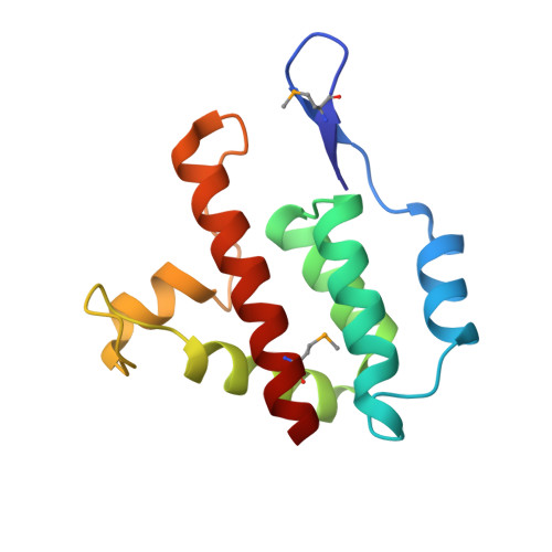
High-resolution structure of a retroviral capsid hexameric amino-terminal domain.

Mortuza, G.B.Haire, L.F.Stevens, A.Smerdon, S.J.Stoye, J.P.Taylor, I.A.

(2004) Nature 431: 481-485

  • DOI: https://doi.org/10.1038/nature02915
  • Primary Citation Related Structures: 
    1U7K

  • PubMed Abstract: 

    Retroviruses are the aetiological agents of a range of human diseases including AIDS and T-cell leukaemias. They follow complex life cycles, which are still only partly understood at the molecular level. Maturation of newly formed retroviral particles is an essential step in production of infectious virions, and requires proteolytic cleavage of Gag polyproteins in the immature particle to form the matrix, capsid and nucleocapsid proteins present in the mature virion. Capsid proteins associate to form a dense viral core that may be spherical, cylindrical or conical depending on the genus of the virus. Nonetheless, these assemblies all appear to be composed of a lattice formed from hexagonal rings, each containing six capsid monomers. Here, we describe the X-ray structure of an individual hexagonal assembly from N-tropic murine leukaemia virus (N-MLV). The interface between capsid monomers is generally polar, consistent with weak interactions within the hexamer. Similar architectures are probably crucial for the regulation of capsid assembly and disassembly in all retroviruses. Together, these observations provide new insights into retroviral uncoating and how cellular restriction factors may interfere with viral replication.


  • Organizational Affiliation
    • Division of Protein Structure, National Institute for Medical Research, The Ridgeway, Mill Hill, London NW7 1AA, UK.

Macromolecule Content 

  • Total Structure Weight: 90.03 kDa 
  • Atom Count: 6,731 
  • Modeled Residue Count: 786 
  • Deposited Residue Count: 786 
  • Unique protein chains: 1

Macromolecules

Find similar proteins by:|  3D Structure
Entity ID: 1
MoleculeChains  Sequence LengthOrganismDetailsImage
Gag polyprotein
A, B, C, D, E
A, B, C, D, E, F
131AKR (endogenous) murine leukemia virusMutation(s): 0 
Gene Names: GAG
UniProt
Find proteins for P03336 (AKV murine leukemia virus)
Explore P03336 
Go to UniProtKB:  P03336
Entity Groups
Sequence Clusters30% Identity50% Identity70% Identity90% Identity95% Identity100% Identity
UniProt GroupP03336
Sequence Annotations
Expand
Reference Sequence

Small Molecules

Modified Residues  1 Unique
IDChains TypeFormula2D DiagramParent
MSE
Query on MSE
A, B, C, D, E
A, B, C, D, E, F
L-PEPTIDE LINKINGC5 H11 N O2 SeMET

Experimental Data & Validation

Experimental Data

  • Method: X-RAY DIFFRACTION
  • Resolution: 1.85 Å
  • R-Value Free:  0.242 (Depositor) 
  • R-Value Work:  0.210 (Depositor) 
  • R-Value Observed: 0.211 (Depositor) 
Space Group: P 1 21 1
Unit Cell:
Length ( Å )Angle ( ˚ )
a = 85.995α = 90
b = 78.34β = 118.86
c = 85.766γ = 90
Software Package:
Software NamePurpose
REFMACrefinement
DENZOdata reduction
SCALEPACKdata scaling
SOLVEphasing

Structure Validation

View Full Validation Report


Entry History 

Deposition Data

Revision History  (Full details and data files)

  • Version 1.0: 2004-10-05
    Type: Initial release
  • Version 1.1: 2008-04-30
    Changes: Version format compliance
  • Version 1.2: 2011-07-13
    Changes: Version format compliance
  • Version 1.3: 2024-11-20
    Changes: Data collection, Database references, Derived calculations, Structure summary