1U4E | pdb_00001u4e

Crystal Structure of Cytoplasmic Domains of GIRK1 channel


Experimental Data Snapshot

  • Method: X-RAY DIFFRACTION
  • Resolution: 2.09 Å
  • R-Value Free: 
    0.235 (Depositor), 0.214 (DCC) 
  • R-Value Work: 
    0.194 (Depositor) 
  • R-Value Observed: 
    0.196 (Depositor) 

Starting Model: experimental
View more details

wwPDB Validation 3D Report Full Report

Validation slider image for 1U4E

This is version 1.4 of the entry. See complete history

Literature

Cytoplasmic domain structures of Kir2.1 and Kir3.1 show sites for modulating gating and rectification

Pegan, S.Arrabit, C.Zhou, W.Kwiatkowski, W.Collins, A.Slesinger, P.A.Choe, S.

(2005) Nat Neurosci 8: 279-287

  • DOI: https://doi.org/10.1038/nn1411
  • Primary Citation Related Structures: 
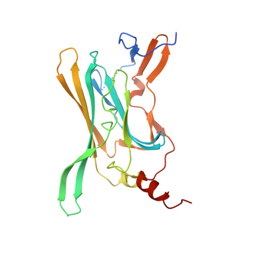
    1U4E, 1U4F

  • PubMed Abstract: 

    N- and C-terminal cytoplasmic domains of inwardly rectifying K (Kir) channels control the ion-permeation pathway through diverse interactions with small molecules and protein ligands in the cytoplasm. Two new crystal structures of the cytoplasmic domains of Kir2.1 (Kir2.1(L)) and the G protein-sensitive Kir3.1 (Kir3.1(S)) channels in the absence of PIP(2) show the cytoplasmic ion-permeation pathways occluded by four cytoplasmic loops that form a girdle around the central pore (G-loop). Significant flexibility of the pore-facing G-loop of Kir2.1(L) and Kir3.1(S) suggests a possible role as a diffusion barrier between cytoplasmic and transmembrane pores. Consistent with this, mutations of the G-loop disrupted gating or inward rectification. Structural comparison shows a di-aspartate cluster on the distal end of the cytoplasmic pore of Kir2.1(L) that is important for modulating inward rectification. Taken together, these results suggest the cytoplasmic domains of Kir channels undergo structural changes to modulate gating and inward rectification.


  • Organizational Affiliation
    • Structural Biology, The Salk Institute, La Jolla, California 92037, USA.

Macromolecule Content 

  • Total Structure Weight: 24.15 kDa 
  • Atom Count: 1,728 
  • Modeled Residue Count: 200 
  • Deposited Residue Count: 212 
  • Unique protein chains: 1

Macromolecules

Find similar proteins by:|  3D Structure
Entity ID: 1
MoleculeChains  Sequence LengthOrganismDetailsImage
G protein-activated inward rectifier potassium channel 1212Mus musculusMutation(s): 0 
Gene Names: Kcnj3GIRK1Kga
UniProt & NIH Common Fund Data Resources
Find proteins for P63250 (Mus musculus)
Explore P63250 
Go to UniProtKB:  P63250
IMPC:  MGI:104742
Entity Groups
Sequence Clusters30% Identity50% Identity70% Identity90% Identity95% Identity100% Identity
UniProt GroupP63250
Sequence Annotations
Expand
Reference Sequence

Experimental Data & Validation

Experimental Data

  • Method: X-RAY DIFFRACTION
  • Resolution: 2.09 Å
  • R-Value Free:  0.235 (Depositor), 0.214 (DCC) 
  • R-Value Work:  0.194 (Depositor) 
  • R-Value Observed: 0.196 (Depositor) 
Space Group: P 4 21 2
Unit Cell:
Length ( Å )Angle ( ˚ )
a = 76.357α = 90
b = 76.357β = 90
c = 92.164γ = 90
Software Package:
Software NamePurpose
REFMACrefinement
HKL-2000data reduction
SCALEPACKdata scaling
AMoREphasing

Structure Validation

View Full Validation Report


Entry History 

Deposition Data

Revision History  (Full details and data files)

  • Version 1.0: 2005-03-08
    Type: Initial release
  • Version 1.1: 2008-04-30
    Changes: Version format compliance
  • Version 1.2: 2011-07-13
    Changes: Derived calculations, Version format compliance
  • Version 1.3: 2017-08-23
    Changes: Source and taxonomy
  • Version 1.4: 2023-08-23
    Changes: Data collection, Database references, Refinement description