1TZD

CRYSTAL STRUCTURE OF THE CATALYTIC CORE OF INOSITOL 1,4,5-TRISPHOSPHATE 3-KINASE


Experimental Data Snapshot

  • Method: X-RAY DIFFRACTION
  • Resolution: 2.20 Å
  • R-Value Free: 0.305 
  • R-Value Work: 0.257 
  • R-Value Observed: 0.260 

wwPDB Validation   3D Report Full Report


Ligand Structure Quality Assessment 


This is version 1.4 of the entry. See complete history


Literature

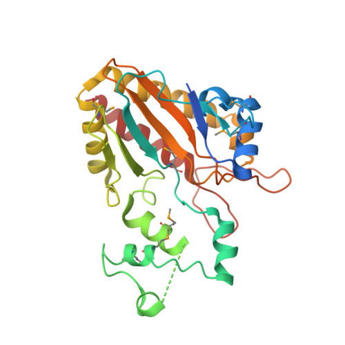
Crystal structure of the catalytic core of inositol 1,4,5-trisphosphate 3-kinase

Miller, G.J.Hurley, J.H.

(2004) Mol Cell 15: 703-711

  • DOI: https://doi.org/10.1016/j.molcel.2004.08.005
  • Primary Citation of Related Structures:  
    1TZD

  • PubMed Abstract: 

    Soluble inositol polyphosphates are ubiquitous second messengers in eukaryotes, and their levels are regulated by an array of specialized kinases. The structure of an archetypal member of this class, inositol 1,4,5-trisphosphate 3-kinase (IP3K), has been determined at 2.2 angstroms resolution in complex with magnesium and adenosine diphosphate. IP3K contains a catalytic domain that is a variant of the protein kinase superfamily, and a novel four-helix substrate binding domain. The two domains are in an open conformation with respect to each other, suggesting that substrate recognition and catalysis by IP3K involves a dynamic conformational cycle. The unique helical domain of IP3K blocks access to the active site by membrane-bound phosphoinositides, explaining the structural basis for soluble inositol polyphosphate specificity.


  • Organizational Affiliation

    Laboratory of Molecular Biology, National Institute of Diabetes and Digestive and Kidney Diseases, National Institutes of Health, U.S. Department of Health and Human Services, Bethesda, MD 20892 USA.


Macromolecules
Find similar proteins by:  (by identity cutoff)  |  3D Structure
Entity ID: 1
MoleculeChains Sequence LengthOrganismDetailsImage
Inositol-trisphosphate 3-kinase A
A, B
275Rattus norvegicusMutation(s): 2 
Gene Names: Itpka
EC: 2.7.1.127
UniProt
Find proteins for P17105 (Rattus norvegicus)
Explore P17105 
Go to UniProtKB:  P17105
Entity Groups  
Sequence Clusters30% Identity50% Identity70% Identity90% Identity95% Identity100% Identity
UniProt GroupP17105
Sequence Annotations
Expand
  • Reference Sequence
Small Molecules
Modified Residues  1 Unique
IDChains TypeFormula2D DiagramParent
MSE
Query on MSE
A, B
L-PEPTIDE LINKINGC5 H11 N O2 SeMET
Experimental Data & Validation

Experimental Data

  • Method: X-RAY DIFFRACTION
  • Resolution: 2.20 Å
  • R-Value Free: 0.305 
  • R-Value Work: 0.257 
  • R-Value Observed: 0.260 
  • Space Group: C 2 2 21
Unit Cell:
Length ( Å )Angle ( ˚ )
a = 71.679α = 90
b = 98.535β = 90
c = 188.745γ = 90
Software Package:
Software NamePurpose
REFMACrefinement
HKL-2000data reduction
SCALEPACKdata scaling
SOLVEphasing

Structure Validation

View Full Validation Report


Ligand Structure Quality Assessment 


Entry History 

Deposition Data

Revision History  (Full details and data files)

  • Version 1.0: 2004-09-21
    Type: Initial release
  • Version 1.1: 2008-04-30
    Changes: Version format compliance
  • Version 1.2: 2011-07-13
    Changes: Advisory, Refinement description, Version format compliance
  • Version 1.3: 2013-06-19
    Changes: Refinement description
  • Version 1.4: 2021-10-27
    Changes: Database references, Derived calculations