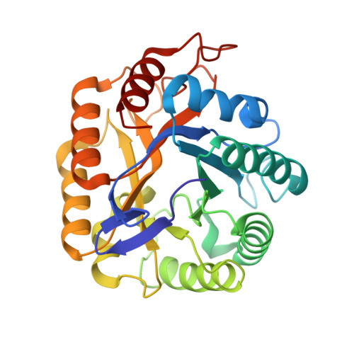
1TVP

Endoglucanase cel5G from Pseudoalteromonas haloplanktis in complex with cellobiose


Experimental Data Snapshot

  • Method: X-RAY DIFFRACTION
  • Resolution: 1.60 Å
  • R-Value Free: 0.189 
  • R-Value Work: 0.158 
  • R-Value Observed: 0.159 

Starting Model: experimental
View more details

wwPDB Validation   3D Report Full Report


This is version 2.1 of the entry. See complete history


Literature

Structure of a Full Length Psychrophilic Cellulase from Pseudoalteromonas haloplanktis revealed by X-ray Diffraction and Small Angle X-ray Scattering

Violot, S.Aghajari, N.Czjzek, M.Feller, G.Sonan, G.K.Gouet, P.Gerday, C.Haser, R.Receveur-Brechot, V.

(2005) J Mol Biol 348: 1211-1224

  • DOI: https://doi.org/10.1016/j.jmb.2005.03.026
  • Primary Citation of Related Structures:  
    1TVN, 1TVP

  • PubMed Abstract: 

    Pseudoalteromonas haloplanktis is a psychrophilic Gram-negative bacterium isolated in Antarctica, that lives on organic remains of algae. This bacterium converts the cellulose, highly constitutive of algae, into an immediate nutritive form by biodegrading this biopolymer. To understand the mechanisms of cold adaptation of its enzymatic components, we studied the structural properties of an endoglucanase, Cel5G, by complementary methods, X-ray crystallography and small angle X-ray scattering. Using X-ray crystallography, we determined the structure of the catalytic core module of this family 5 endoglucanase, at 1.4A resolution in its native form and at 1.6A in the cellobiose-bound form. The catalytic module of Cel5G presents the (beta/alpha)(8)-barrel structure typical of clan GH-A of glycoside hydrolase families. The structural comparison of the catalytic core of Cel5G with the mesophilic catalytic core of Cel5A from Erwinia chrysanthemi revealed modifications at the atomic level leading to higher flexibility and thermolability, which might account for the higher activity of Cel5G at low temperatures. Using small angle X-ray scattering we further explored the structure at the entire enzyme level. We analyzed the dimensions, shape, and conformation of Cel5G full length in solution and especially of the linker between the catalytic module and the cellulose-binding module. The results showed that the linker is unstructured, and unusually long and flexible, a peculiarity that distinguishes it from its mesophilic counterpart. Loops formed at the base by disulfide bridges presumably add constraints to stabilize the most extended conformations. These results suggest that the linker plays a major role in cold adaptation of this psychrophilic enzyme, allowing steric optimization of substrate accessibility.


  • Organizational Affiliation

    Laboratoire de BioCristallographie, Institut de Biologie et Chimie des Protéines, CNRS et Université Claude Bernard Lyon 1, UMR 5086, IFR 128 Biosciences Lyon-Gerland, 7 Passage du Vercors, F-69367 Lyon Cedex 07, France.


Macromolecules
Find similar proteins by:  (by identity cutoff)  |  3D Structure
Entity ID: 1
MoleculeChains Sequence LengthOrganismDetailsImage
cellulase
A, B
293Pseudoalteromonas haloplanktisMutation(s): 0 
Gene Names: celG
EC: 3.2.1.4
UniProt
Find proteins for O86099 (Pseudoalteromonas haloplanktis)
Explore O86099 
Go to UniProtKB:  O86099
Entity Groups  
Sequence Clusters30% Identity50% Identity70% Identity90% Identity95% Identity100% Identity
UniProt GroupO86099
Sequence Annotations
Expand
  • Reference Sequence
Oligosaccharides

Help

Entity ID: 2
MoleculeChains Length2D Diagram Glycosylation3D Interactions
beta-D-glucopyranose-(1-4)-beta-D-glucopyranose
C
2N/A
Glycosylation Resources
GlyTouCan:  G84824ZO
GlyCosmos:  G84824ZO
Small Molecules
Ligands 1 Unique
IDChains Name / Formula / InChI Key2D Diagram3D Interactions
EPE
Query on EPE

Download Ideal Coordinates CCD File 
D [auth A]4-(2-HYDROXYETHYL)-1-PIPERAZINE ETHANESULFONIC ACID
C8 H18 N2 O4 S
JKMHFZQWWAIEOD-UHFFFAOYSA-N
Biologically Interesting Molecules (External Reference) 1 Unique
Entity ID: 2
IDChains NameType/Class2D Diagram3D Interactions
PRD_900005
Query on PRD_900005
C
beta-cellobioseOligosaccharide / Metabolism
Experimental Data & Validation

Experimental Data

  • Method: X-RAY DIFFRACTION
  • Resolution: 1.60 Å
  • R-Value Free: 0.189 
  • R-Value Work: 0.158 
  • R-Value Observed: 0.159 
  • Space Group: P 21 21 21
Unit Cell:
Length ( Å )Angle ( ˚ )
a = 135.5α = 90
b = 78.9β = 90
c = 44γ = 90
Software Package:
Software NamePurpose
CNSrefinement
MOSFLMdata reduction
CCP4data scaling
AMoREphasing

Structure Validation

View Full Validation Report


Entry History 

Deposition Data

Revision History  (Full details and data files)

  • Version 1.0: 2005-05-17
    Type: Initial release
  • Version 1.1: 2008-04-30
    Changes: Version format compliance
  • Version 1.2: 2011-07-13
    Changes: Version format compliance
  • Version 2.0: 2020-07-29
    Type: Remediation
    Reason: Carbohydrate remediation
    Changes: Atomic model, Data collection, Derived calculations, Non-polymer description, Structure summary
  • Version 2.1: 2023-10-25
    Changes: Data collection, Database references, Refinement description, Structure summary