1TTT

Phe-tRNA, elongation factoR EF-TU:GDPNP ternary complex


Experimental Data Snapshot

  • Method: X-RAY DIFFRACTION
  • Resolution: 2.70 Å
  • R-Value Free: 0.290 
  • R-Value Work: 0.206 

wwPDB Validation   3D Report Full Report


This is version 1.3 of the entry. See complete history


Literature

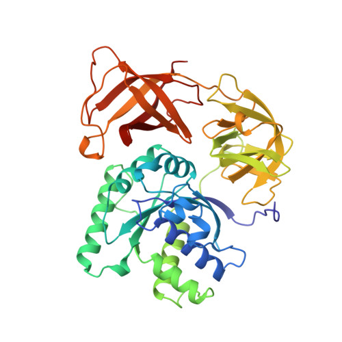
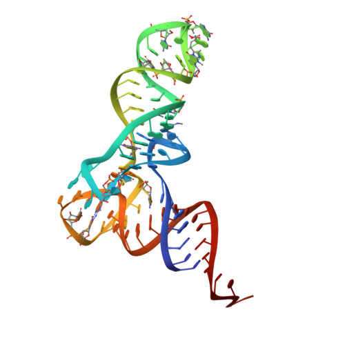
Crystal structure of the ternary complex of Phe-tRNAPhe, EF-Tu, and a GTP analog.

Nissen, P.Kjeldgaard, M.Thirup, S.Polekhina, G.Reshetnikova, L.Clark, B.F.Nyborg, J.

(1995) Science 270: 1464-1472

  • DOI: https://doi.org/10.1126/science.270.5241.1464
  • Primary Citation of Related Structures:  
    1TTT

  • PubMed Abstract: 

    The structure of the ternary complex consisting of yeast phenylalanyl-transfer RNA (Phe-tRNAPhe), Thermus aquaticus elongation factor Tu (EF-Tu), and the guanosine triphosphate (GTP) analog GDPNP was determined by x-ray crystallography at 2.7 angstrom resolution. The ternary complex participates in placing the amino acids in their correct order when messenger RNA is translated into a protein sequence on the ribosome. The EF-Tu-GDPNP component binds to one side of the acceptor helix of Phe-tRNAPhe involving all three domains of EF-Tu. Binding sites for the phenylalanylated CCA end and the phosphorylated 5' end are located at domain interfaces, whereas the T stem interacts with the surface of the beta-barrel domain 3. The binding involves many conserved residues in EF-Tu. The overall shape of the ternary complex is similar to that of the translocation factor, EF-G-GDP, and this suggests a novel mechanism involving "molecular mimicry" in the translational apparatus.


  • Organizational Affiliation

    Department of Biostructural Chemistry, Institute of Chemistry, Aarhus University, Denmark.


Macromolecules

Find similar proteins by:  (by identity cutoff)  |  3D Structure
Entity ID: 2
MoleculeChains Sequence LengthOrganismDetailsImage
OF ELONGATION FACTOR TU (EF-TU)D [auth A],
E [auth B],
F [auth C]
405Thermus aquaticusMutation(s): 0 
UniProt
Find proteins for Q01698 (Thermus aquaticus)
Explore Q01698 
Go to UniProtKB:  Q01698
Entity Groups  
Sequence Clusters30% Identity50% Identity70% Identity90% Identity95% Identity100% Identity
UniProt GroupQ01698
Sequence Annotations
Expand
  • Reference Sequence
Find similar nucleic acids by:  (by identity cutoff)  |  3D Structure
Entity ID: 1
MoleculeChains LengthOrganismImage
TRANSFER RIBONUCLEIC ACID (YEAST, PHE)A [auth D],
B [auth E],
C [auth F]
76N/A
Sequence Annotations
Expand
  • Reference Sequence
Small Molecules
Ligands 3 Unique
IDChains Name / Formula / InChI Key2D Diagram3D Interactions
GNP
Query on GNP

Download Ideal Coordinates CCD File 
N [auth A],
P [auth B],
R [auth C]
PHOSPHOAMINOPHOSPHONIC ACID-GUANYLATE ESTER
C10 H17 N6 O13 P3
UQABYHGXWYXDTK-UUOKFMHZSA-N
PHE
Query on PHE

Download Ideal Coordinates CCD File 
H [auth D],
J [auth E],
L [auth F]
PHENYLALANINE
C9 H11 N O2
COLNVLDHVKWLRT-QMMMGPOBSA-N
MG
Query on MG

Download Ideal Coordinates CCD File 
G [auth D]
I [auth E]
K [auth F]
M [auth A]
O [auth B]
G [auth D],
I [auth E],
K [auth F],
M [auth A],
O [auth B],
Q [auth C]
MAGNESIUM ION
Mg
JLVVSXFLKOJNIY-UHFFFAOYSA-N
Experimental Data & Validation

Experimental Data

  • Method: X-RAY DIFFRACTION
  • Resolution: 2.70 Å
  • R-Value Free: 0.290 
  • R-Value Work: 0.206 
  • Space Group: C 1 2 1
Unit Cell:
Length ( Å )Angle ( ˚ )
a = 206.835α = 90
b = 122.354β = 126.3
c = 151.552γ = 90
Software Package:
Software NamePurpose
TNTrefinement
DENZOdata reduction

Structure Validation

View Full Validation Report


Entry History 

Deposition Data

Revision History  (Full details and data files)

  • Version 1.0: 1996-12-23
    Type: Initial release
  • Version 1.1: 2008-05-22
    Changes: Version format compliance
  • Version 1.2: 2011-07-13
    Changes: Non-polymer description, Version format compliance
  • Version 1.3: 2024-02-14
    Changes: Data collection, Database references, Derived calculations, Other