1TRB | pdb_00001trb

CONVERGENT EVOLUTION OF SIMILAR FUNCTION IN TWO STRUCTURALLY DIVERGENT ENZYMES


Experimental Data Snapshot

  • Method: X-RAY DIFFRACTION
  • Resolution: 2.00 Å
  • R-Value Work: 
    0.177 (Depositor), 0.174 (DCC) 
  • R-Value Observed: 
    0.177 (Depositor) 

wwPDB Validation 3D Report Full Report

Validation slider image for 1TRB

Ligand Structure Quality Assessment 


This is version 1.3 of the entry. See complete history

Literature

Convergent evolution of similar function in two structurally divergent enzymes.

Kuriyan, J.Krishna, T.S.Wong, L.Guenther, B.Pahler, A.Williams Jr., C.H.Model, P.

(1991) Nature 352: 172-174

  • DOI: https://doi.org/10.1038/352172a0
  • Primary Citation Related Structures: 
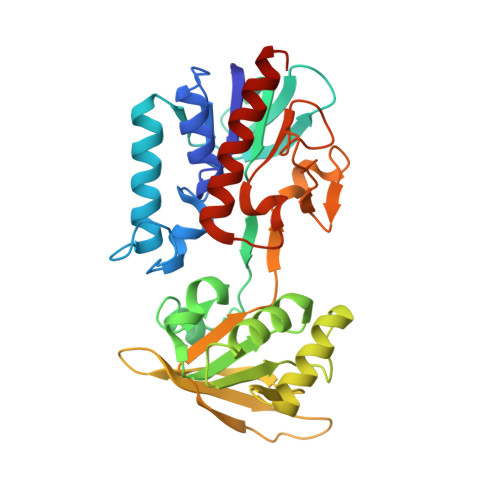
    1TRB

  • PubMed Abstract: 

    An example of two related enzymes that catalyse similar reactions but possess different active sites is provided by comparing the structure of Escherichia coli thioredoxin reductase with glutathione reductase. Both are dimeric enzymes that catalyse the reduction of disulphides by pyridine nucleotides through an enzyme disulphide and a flavin. Human glutathione reductase contains four structural domains within each molecule: the flavin-adenine dinucleotide (FAD)- and nicotinamide-adenine dinucleotide phosphate (NADPH)-binding domains, the 'central' domain and the C-terminal domain that provides the dimer interface and part of the active site. Although both enzymes share the same catalytic mechanism and similar tertiary structures, their active sites do not resemble each other. We have determined the crystal structure of E. coli thioredoxin reductase at 2 A resolution, and show that thioredoxin reductase lacks the domain that provides the dimer interface in glutathione reductase, and forms a completely different dimeric structure. The catalytically active disulphides are located in different domains on opposite sides of the flavin ring system. This suggests that these enzymes diverged from an ancestral nucleotide-binding protein and acquired their disulphide reductase activities independently.


  • Organizational Affiliation
    • Rockefeller University, New York 10021.

Macromolecule Content 

  • Total Structure Weight: 35.3 kDa 
  • Atom Count: 2,611 
  • Modeled Residue Count: 316 
  • Deposited Residue Count: 320 
  • Unique protein chains: 1

Macromolecules

Find similar proteins by:|  3D Structure
Entity ID: 1
MoleculeChains  Sequence LengthOrganismDetailsImage
THIOREDOXIN REDUCTASE320Escherichia coliMutation(s): 0 
EC: 1.6.4.5 (PDB Primary Data), 1.8.1.9 (UniProt)
UniProt
Find proteins for P0A9P4 (Escherichia coli (strain K12))
Explore P0A9P4 
Go to UniProtKB:  P0A9P4
Entity Groups
Sequence Clusters30% Identity50% Identity70% Identity90% Identity95% Identity100% Identity
UniProt GroupP0A9P4
Sequence Annotations
Expand
Reference Sequence

Small Molecules

Ligands 1 Unique
IDChains Name / Formula / InChI Key2D Diagram3D Interactions
FAD

Query on FAD


Download:Ideal Coordinates CCD File
B [auth A]FLAVIN-ADENINE DINUCLEOTIDE
C27 H33 N9 O15 P2
VWWQXMAJTJZDQX-UYBVJOGSSA-N

Experimental Data & Validation

Experimental Data

  • Method: X-RAY DIFFRACTION
  • Resolution: 2.00 Å
  • R-Value Work:  0.177 (Depositor), 0.174 (DCC) 
  • R-Value Observed: 0.177 (Depositor) 
Space Group: P 63 2 2
Unit Cell:
Length ( Å )Angle ( ˚ )
a = 123.1α = 90
b = 123.1β = 90
c = 81.4γ = 120
Software Package:
Software NamePurpose
X-PLORmodel building
X-PLORrefinement
X-PLORphasing

Structure Validation

View Full Validation Report


Ligand Structure Quality Assessment 


Entry History 

Deposition Data

Revision History  (Full details and data files)

  • Version 1.0: 1994-01-31
    Type: Initial release
  • Version 1.1: 2008-03-24
    Changes: Version format compliance
  • Version 1.2: 2011-07-13
    Changes: Derived calculations, Version format compliance
  • Version 1.3: 2024-02-14
    Changes: Data collection, Database references, Derived calculations, Other