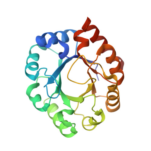
1TQX | pdb_00001tqx

Crystal Structure of Pfal009167 A Putative D-Ribulose 5-Phosphate 3-Epimerase from P.falciparum


Experimental Data Snapshot

  • Method: X-RAY DIFFRACTION
  • Resolution: 2.00 Å
  • R-Value Free: 
    0.233 (Depositor), 0.215 (DCC) 
  • R-Value Work: 
    0.212 (Depositor), 0.215 (DCC) 
  • R-Value Observed: 
    0.212 (Depositor) 

Starting Model: experimental
View more details

wwPDB Validation 3D Report Full Report

Validation slider image for 1TQX

This is version 1.3 of the entry. See complete history. 

Literature

Structure of a ribulose 5-phosphate 3-epimerase from Plasmodium falciparum.

Caruthers, J., Bosch, J., Buckner, F., Van Voorhis, W., Myler, P., Worthey, E., Mehlin, C., Boni, E., DeTitta, G., Luft, J., Lauricella, A., Kalyuzhniy, O., Anderson, L., Zucker, F., Soltis, M., Hol, W.G.

(2006) Proteins 62: 338-342

  • DOI: https://doi.org/10.1002/prot.20764
  • Primary Citation Related Structures: 
    1TQX

  • PubMed Abstract: 

    The crystal structure of Pfal009167AAA, a putative ribulose 5-phosphate 3-epimerase (PfalRPE) from Plasmodium falciparum, has been determined to 2 A resolution. RPE represents an exciting potential drug target for developing antimalarials because it is involved in the shikimate and the pentose phosphate pathways. The structure is a classic TIM-barrel fold. A coordinated Zn ion and a bound sulfate ion in the active site of the enzyme allow for a greater understanding of the mechanism of action of this enzyme. This structure is solved in the framework of the Structural Genomics of Pathogenic Protozoa (SGPP) consortium.


  • Organizational Affiliation: 
    • SLAC, Stanford University, Menlo Park, California, USA.

Macromolecule Content 

  • Total Structure Weight: 51.48 kDa 
  • Atom Count: 3,752 
  • Modeled Residue Count: 442 
  • Deposited Residue Count: 454 
  • Unique protein chains: 1

Macromolecules

Find similar proteins by:|  3D Structure
Entity ID: 1
MoleculeChains  Sequence LengthOrganismDetailsImage
D-ribulose-5-phosphate 3-epimerase, putative
A, B
227Plasmodium falciparum 3D7Mutation(s): 0 
EC: 5.1.3.1
UniProt
Find proteins for Q8I5L3 (Plasmodium falciparum (isolate 3D7))
Explore Q8I5L3 
Go to UniProtKB:  Q8I5L3
Entity Groups
Sequence Clusters30% Identity50% Identity70% Identity90% Identity95% Identity100% Identity
UniProt GroupQ8I5L3
Sequence Annotations
Expand
Reference Sequence

Experimental Data & Validation

Experimental Data

  • Method: X-RAY DIFFRACTION
  • Resolution: 2.00 Å
  • R-Value Free:  0.233 (Depositor), 0.215 (DCC) 
  • R-Value Work:  0.212 (Depositor), 0.215 (DCC) 
  • R-Value Observed: 0.212 (Depositor) 
Space Group: P 21 21 21
Unit Cell:
Length ( Å )Angle ( ˚ )
a = 79.415α = 90
b = 84.454β = 90
c = 120.717γ = 90
Software Package:
Software NamePurpose
CNSrefinement
DENZOdata reduction
SCALEPACKdata scaling
EPMRphasing

Structure Validation

View Full Validation Report


Entry History 

Deposition Data

Revision History  (Full details and data files)

  • Version 1.0: 2004-12-21
    Type: Initial release
  • Version 1.1: 2008-04-30
    Changes: Version format compliance
  • Version 1.2: 2011-07-13
    Changes: Derived calculations, Source and taxonomy, Version format compliance
  • Version 1.3: 2023-08-23
    Changes: Data collection, Database references, Derived calculations, Refinement description