1TON | pdb_00001ton

RAT SUBMAXILLARY GLAND SERINE PROTEASE, TONIN. STRUCTURE SOLUTION AND REFINEMENT AT 1.8 ANGSTROMS RESOLUTION


Experimental Data Snapshot

  • Method: X-RAY DIFFRACTION
  • Resolution: 1.80 Å
  • R-Value Observed: 
    0.196 (Depositor) 

wwPDB Validation 3D Report Full Report

Validation slider image for 1TON

This is version 1.4 of the entry. See complete history

Literature

Rat submaxillary gland serine protease, tonin. Structure solution and refinement at 1.8 A resolution.

Fujinaga, M.James, M.N.

(1987) J Mol Biology 195: 373-396

  • DOI: https://doi.org/10.1016/0022-2836(87)90658-9
  • Primary Citation Related Structures: 
    1TON

  • PubMed Abstract: 

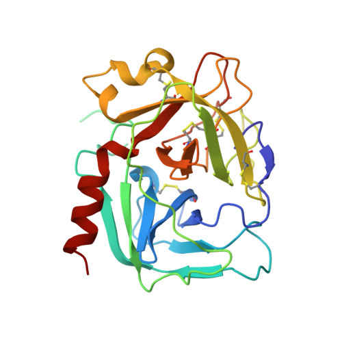
    Tonin is a mammalian serine protease that is capable of generating the vasoconstrictive agent, angiotensin II, directly from its precursor protein, angiotensinogen, a process that normally requires two enzymes, renin and angiotensin-converting enzyme. The X-ray crystallographic structure determination and refinement of tonin at 1.8 A resolution and the analysis of the resulting model are reported. The initial phases were obtained by the method of molecular replacement using as the search model the structure of bovine trypsin. The refined model of tonin consists of 227 amino acid residues out of the 235 in the complete molecule, 149 water molecules, and one zinc ion. The R-factor (R = sigma Fo - Fc/sigma Fo) is 0.196 for the 14,997 measured data between 8 and 1.8 A resolution with I greater than or equal to sigma (I). It is estimated that the overall root-mean-square error in the coordinates is about 0.3 A. The structure of tonin that has been determined is not in its active conformation, but one that has been perturbed by the binding of Zn2+ in the active site. Zn2+ was included in the buffer to aid the crystallization. Nevertheless, the structure of tonin that is described is for the most part similar to its native form as indicated by the close tertiary structural homology with kallikrein. The differences in the structures of the two enzymes are concentrated in several loop regions; these structural differences are probably responsible for the differences in their reactivities and specificities.


  • Organizational Affiliation
    • Department of Biochemistry, University of Alberta, Edmonton, Canada.

Macromolecule Content 

  • Total Structure Weight: 25.75 kDa 
  • Atom Count: 1,884 
  • Modeled Residue Count: 228 
  • Deposited Residue Count: 235 
  • Unique protein chains: 1

Macromolecules

Find similar proteins by:|  3D Structure
Entity ID: 1
MoleculeChains  Sequence LengthOrganismDetailsImage
TONIN235Rattus rattusMutation(s): 0 
EC: 3.4.21.35
UniProt
Find proteins for P00759 (Rattus norvegicus)
Explore P00759 
Go to UniProtKB:  P00759
Entity Groups
Sequence Clusters30% Identity50% Identity70% Identity90% Identity95% Identity100% Identity
UniProt GroupP00759
Sequence Annotations
Expand
Reference Sequence

Small Molecules

Ligands 1 Unique
IDChains Name / Formula / InChI Key2D Diagram3D Interactions
ZN

Query on ZN


Download:Ideal Coordinates CCD File
B [auth A]ZINC ION
Zn
PTFCDOFLOPIGGS-UHFFFAOYSA-N

Experimental Data & Validation

Experimental Data

  • Method: X-RAY DIFFRACTION
  • Resolution: 1.80 Å
  • R-Value Observed: 0.196 (Depositor) 
Space Group: P 43 21 2
Unit Cell:
Length ( Å )Angle ( ˚ )
a = 48.58α = 90
b = 48.58β = 90
c = 200.2γ = 90
Software Package:
Software NamePurpose
PROLSQrefinement

Structure Validation

View Full Validation Report


Entry History 

Deposition Data

Revision History  (Full details and data files)

  • Version 1.0: 1988-01-16
    Type: Initial release
  • Version 1.1: 2008-03-24
    Changes: Version format compliance
  • Version 1.2: 2011-07-13
    Changes: Derived calculations, Version format compliance
  • Version 1.3: 2017-11-29
    Changes: Advisory, Derived calculations, Other
  • Version 1.4: 2024-10-30
    Changes: Advisory, Data collection, Database references, Derived calculations, Structure summary