1TND | pdb_00001tnd

THE 2.2 ANGSTROMS CRYSTAL STRUCTURE OF TRANSDUCIN-ALPHA COMPLEXED WITH GTP GAMMA S


Experimental Data Snapshot

  • Method: X-RAY DIFFRACTION
  • Resolution: 2.20 Å
  • R-Value Work: 
    0.190 (Depositor) 
  • R-Value Observed: 
    0.190 (Depositor) 

wwPDB Validation 3D Report Full Report

Validation slider image for 1TND

This is version 1.4 of the entry. See complete history

Literature

The 2.2 A crystal structure of transducin-alpha complexed with GTP gamma S.

Noel, J.P.Hamm, H.E.Sigler, P.B.

(1993) Nature 366: 654-663

  • DOI: https://doi.org/10.1038/366654a0
  • Primary Citation Related Structures: 
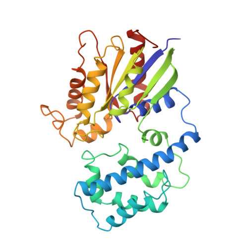
    1TND

  • PubMed Abstract: 

    The 2.2 A crystal structure of activated rod transducin, Gt alpha.GTP gamma S, shows the bound GTP gamma S molecule occluded deep in a cleft between a domain structurally homologous to small GTPases and a helical domain unique to heterotrimeric G proteins. The structure, when combined with biochemical and genetic studies, suggests: how an activated receptor might open this cleft to allow nucleotide exchange; a mechanism for GTP-induced changes in effector and receptor binding surfaces; and a mechanism for GTPase activity not evident from previous data.


  • Organizational Affiliation
    • Department of Molecular Biophysics and Biochemistry, Yale University, New Haven, Connecticut 06510.

Macromolecule Content 

  • Total Structure Weight: 113.94 kDa 
  • Atom Count: 8,599 
  • Modeled Residue Count: 955 
  • Deposited Residue Count: 972 
  • Unique protein chains: 1

Macromolecules

Find similar proteins by:|  3D Structure
Entity ID: 1
MoleculeChains  Sequence LengthOrganismDetailsImage
TRANSDUCIN
A, B, C
324Bos taurusMutation(s): 0 
UniProt
Find proteins for P04695 (Bos taurus)
Explore P04695 
Go to UniProtKB:  P04695
Entity Groups
Sequence Clusters30% Identity50% Identity70% Identity90% Identity95% Identity100% Identity
UniProt GroupP04695
Sequence Annotations
Expand
Reference Sequence

Small Molecules

Ligands 3 Unique
IDChains Name / Formula / InChI Key2D Diagram3D Interactions
GSP

Query on GSP


Download:Ideal Coordinates CCD File
G [auth A],
K [auth B],
O [auth C]
5'-GUANOSINE-DIPHOSPHATE-MONOTHIOPHOSPHATE
C10 H16 N5 O13 P3 S
XOFLBQFBSOEHOG-UUOKFMHZSA-N
CAC

Query on CAC


Download:Ideal Coordinates CCD File
E [auth A]
F [auth A]
I [auth B]
J [auth B]
M [auth C]
E [auth A],
F [auth A],
I [auth B],
J [auth B],
M [auth C],
N [auth C]
CACODYLATE ION
C2 H6 As O2
OGGXGZAMXPVRFZ-UHFFFAOYSA-M
MG

Query on MG


Download:Ideal Coordinates CCD File
D [auth A],
H [auth B],
L [auth C]
MAGNESIUM ION
Mg
JLVVSXFLKOJNIY-UHFFFAOYSA-N

Experimental Data & Validation

Experimental Data

  • Method: X-RAY DIFFRACTION
  • Resolution: 2.20 Å
  • R-Value Work:  0.190 (Depositor) 
  • R-Value Observed: 0.190 (Depositor) 
Space Group: P 1 21 1
Unit Cell:
Length ( Å )Angle ( ˚ )
a = 74.5α = 90
b = 108.3β = 112.3
c = 79γ = 90
Software Package:
Software NamePurpose
X-PLORmodel building
X-PLORrefinement
X-PLORphasing

Structure Validation

View Full Validation Report


Entry History 

Deposition Data

Revision History  (Full details and data files)

  • Version 1.0: 1994-07-31
    Type: Initial release
  • Version 1.1: 2008-03-24
    Changes: Version format compliance
  • Version 1.2: 2011-07-13
    Changes: Non-polymer description, Version format compliance
  • Version 1.3: 2017-11-29
    Changes: Derived calculations, Other
  • Version 1.4: 2024-02-14
    Changes: Data collection, Database references, Derived calculations