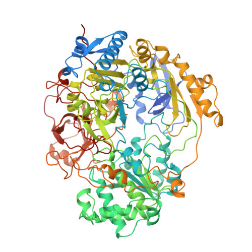
1TMO | pdb_00001tmo

TRIMETHYLAMINE N-OXIDE REDUCTASE FROM SHEWANELLA MASSILIA


Experimental Data Snapshot

  • Method: X-RAY DIFFRACTION
  • Resolution: 2.50 Å
  • R-Value Free: 
    0.247 (Depositor) 
  • R-Value Work: 
    0.182 (Depositor), 0.209 (DCC) 
  • R-Value Observed: 
    0.182 (Depositor) 

wwPDB Validation 3D Report Full Report

Validation slider image for 1TMO

This is version 2.0 of the entry. See complete history

Literature

Crystal structure of oxidized trimethylamine N-oxide reductase from Shewanella massilia at 2.5 A resolution.

Czjzek, M.Dos Santos, J.P.Pommier, J.Giordano, G.Mejean, V.Haser, R.

(1998) J Mol Biol 284: 435-447

  • DOI: https://doi.org/10.1006/jmbi.1998.2156
  • Primary Citation Related Structures: 
    1TMO

  • PubMed Abstract: 

    The periplasmic trimethylamine N-oxide (TMAO) reductase from the marine bacteria Shewanella massilia is involved in a respiratory chain, having trimethylamine N-oxide as terminal electron acceptor. This molybdoenzyme belongs to the dimethyl sulfoxide (DMSO) reductase family, but has a different substrate specificity than its homologous enzyme. While the DMSO reductases reduce a broad spectra of organic S-oxide and N-oxide compounds, TMAO reductase from Shewanella massilia reduces only TMAO as the natural compound. The crystal structure was solved by molecular replacement with the coordinates of the DMSO reductase from Rhodobacter sphaeroides. The overall fold of the protein structure is essentially the same as the DMSO reductase structures, organized into four domains. The molybdenum coordination sphere is closest to that described in the DMSO reductase of Rhodobacter capsulatus. The structural differences found in the protein environment of the active site could be related to the differences in substrate specificity of these enzymes. In close vicinity of the molybdenum ion a tyrosine residue is missing in the TMAO reductase, leaving a greater space accessible to the solvent. This tyrosine residue has contacts to the oxo groups in the DMSO reductase structures. The arrangement and number of charged residues lining the inner surface of the funnel-like entrance to the active site, is different in the TMAO reductase than in the DMSO reductases from Rhodobacter species. Furthermore a surface loop at the top of the active-site funnel, for which no density was present in the DMSO reductase structures, is well defined in the oxidized form of the TMAO reductase structure, and is located on the border of the funnel-like entrance of the active center.


  • Organizational Affiliation
    • Laboratoire d'Architecture et Fonction de Macromolécules Biologiques, AFMB-CNRS Marseille, IBSM, 31 chemin Joseph Aiguier, Marseille Cedex 20, 13402, France. czjzek@afmb.cnrs-mrs.fr

Macromolecule Content 

  • Total Structure Weight: 94.08 kDa 
  • Atom Count: 6,857 
  • Modeled Residue Count: 794 
  • Deposited Residue Count: 829 
  • Unique protein chains: 1

Macromolecules

Find similar proteins by:|  3D Structure
Entity ID: 1
MoleculeChains  Sequence LengthOrganismDetailsImage
TRIMETHYLAMINE N-OXIDE REDUCTASE829Shewanella massiliaMutation(s): 0 
EC: 1.6.6.9 (PDB Primary Data), 1.7.2.3 (UniProt)
UniProt
Find proteins for O87948 (Shewanella massilia)
Explore O87948 
Go to UniProtKB:  O87948
Entity Groups
Sequence Clusters30% Identity50% Identity70% Identity90% Identity95% Identity100% Identity
UniProt GroupO87948
Sequence Annotations
Expand
Reference Sequence

Small Molecules

Ligands 1 Unique
IDChains Name / Formula / InChI Key2D Diagram3D Interactions
BKG

Query on BKG


Download:Ideal Coordinates CCD File
B [auth A]bis-molybdopterin guanine dinucleotide dioxo molybdenum
C40 H48 Mo N20 O28 P4 S4
BINXIZOEMHRYMB-XBFBQCSYSA-J

Experimental Data & Validation

Experimental Data

  • Method: X-RAY DIFFRACTION
  • Resolution: 2.50 Å
  • R-Value Free:  0.247 (Depositor) 
  • R-Value Work:  0.182 (Depositor), 0.209 (DCC) 
  • R-Value Observed: 0.182 (Depositor) 
Space Group: P 21 21 2
Unit Cell:
Length ( Å )Angle ( ˚ )
a = 123.32α = 90
b = 140.7β = 90
c = 59.39γ = 90
Software Package:
Software NamePurpose
X-PLORmodel building
X-PLORrefinement
X-PLORphasing

Structure Validation

View Full Validation Report


Entry History 

Deposition Data

Revision History  (Full details and data files)

  • Version 1.0: 1999-03-30
    Type: Initial release
  • Version 1.1: 2008-03-24
    Changes: Version format compliance
  • Version 1.2: 2011-07-13
    Changes: Version format compliance
  • Version 1.3: 2024-02-14
    Changes: Data collection, Database references, Derived calculations, Other
  • Version 2.0: 2026-08-12
    Type: Remediation
    Reason: Metalloprotein remediation
    Changes: Atomic model, Data collection, Derived calculations, Non-polymer description, Structure summary