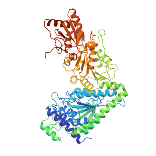
1TKB

SPECIFICITY OF COENZYME BINDING IN THIAMIN DIPHOSPHATE DEPENDENT ENZYMES: CRYSTAL STRUCTURES OF YEAST TRANSKETOLASE IN COMPLEX WITH ANALOGS OF THIAMIN DIPHOSPHATE


Experimental Data Snapshot

  • Method: X-RAY DIFFRACTION
  • Resolution: 2.30 Å
  • R-Value Work: 0.185 
  • R-Value Observed: 0.185 

wwPDB Validation   3D Report Full Report


This is version 1.3 of the entry. See complete history


Literature

Specificity of coenzyme binding in thiamin diphosphate-dependent enzymes. Crystal structures of yeast transketolase in complex with analogs of thiamin diphosphate.

Konig, S.Schellenberger, A.Neef, H.Schneider, G.

(1994) J Biol Chem 269: 10879-10882

  • Primary Citation of Related Structures:  
    1TKA, 1TKB, 1TKC

  • PubMed Abstract: 

    The three-dimensional structures of complexes of yeast apotransketolase with the coenzyme analogs 6'-methyl, N1'-pyridyl, and N3'-pyridyl thiamin diphosphate, respectively, were determined with protein crystallographic methods. All three coenzyme analogs bind to the enzyme in a fashion highly similar to the cofactor thiamin diphosphate. Thus, either one of the hydrogen bonds of the pyrimidine ring nitrogens to the protein is sufficient for proper binding and positioning of the cofactor. The lack of catalytic activity of the N3'-pyridyl analog is not due to incorrect orientation of the pyrimidine ring, but results from the absence of the hydrogen bond between the N1' nitrogen atom and the conserved residue Glu418. The structure analysis provides further evidence for the importance of this conserved interaction for enzymatic thiamin catalysis.


  • Organizational Affiliation

    Department of Molecular Biology, Swedish University of Agricultural Sciences, Uppsala.


Macromolecules
Find similar proteins by:  (by identity cutoff)  |  3D Structure
Entity ID: 1
MoleculeChains Sequence LengthOrganismDetailsImage
TRANSKETOLASE
A, B
678Saccharomyces cerevisiaeMutation(s): 0 
EC: 2.2.1.1
UniProt
Find proteins for P23254 (Saccharomyces cerevisiae (strain ATCC 204508 / S288c))
Explore P23254 
Go to UniProtKB:  P23254
Entity Groups  
Sequence Clusters30% Identity50% Identity70% Identity90% Identity95% Identity100% Identity
UniProt GroupP23254
Sequence Annotations
Expand
  • Reference Sequence
Small Molecules
Binding Affinity Annotations 
IDSourceBinding Affinity
N1T PDBBind:  1TKB Ki: 10 (nM) from 1 assay(s)
Experimental Data & Validation

Experimental Data

  • Method: X-RAY DIFFRACTION
  • Resolution: 2.30 Å
  • R-Value Work: 0.185 
  • R-Value Observed: 0.185 
  • Space Group: P 21 21 21
Unit Cell:
Length ( Å )Angle ( ˚ )
a = 76.3α = 90
b = 113.3β = 90
c = 160.9γ = 90
Software Package:
Software NamePurpose
X-PLORmodel building
X-PLORrefinement
X-PLORphasing

Structure Validation

View Full Validation Report


Entry History 

Deposition Data

Revision History  (Full details and data files)

  • Version 1.0: 1994-11-30
    Type: Initial release
  • Version 1.1: 2008-03-24
    Changes: Version format compliance
  • Version 1.2: 2011-07-13
    Changes: Version format compliance
  • Version 1.3: 2024-02-14
    Changes: Data collection, Database references, Derived calculations, Other