1TJL | pdb_00001tjl

Crystal structure of transcription factor DksA from E. coli


Experimental Data Snapshot

  • Method: X-RAY DIFFRACTION
  • Resolution: 2.00 Å
  • R-Value Free: 
    0.268 (Depositor), 0.264 (DCC) 
  • R-Value Work: 
    0.228 (Depositor), 0.226 (DCC) 
  • R-Value Observed: 
    0.228 (Depositor) 

wwPDB Validation 3D Report Full Report

Validation slider image for 1TJL

This is version 1.4 of the entry. See complete history

Literature

Regulation through the secondary channel--structural framework for ppGpp-DksA synergism during transcription

Perederina, A.Svetlov, V.Vassylyeva, M.N.Tahirov, T.H.Yokoyama, S.Artsimovitch, I.Vassylyev, D.G.

(2004) Cell 118: 297-309

  • DOI: https://doi.org/10.1016/j.cell.2004.06.030
  • Primary Citation Related Structures: 
    1TJL

  • PubMed Abstract: 

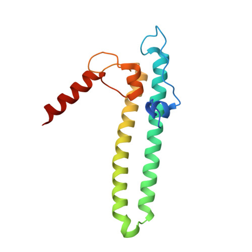
    Bacterial transcription is regulated by the alarmone ppGpp, which binds near the catalytic site of RNA polymerase (RNAP) and modulates its activity. We show that the DksA protein is a crucial component of ppGpp-dependent regulation. The 2.0 A resolution structure of Escherichia coli DksA reveals a globular domain and a coiled coil with two highly conserved Asp residues at its tip that is reminiscent of the transcript cleavage factor GreA. This structural similarity suggests that DksA coiled coil protrudes into the RNAP secondary channel to coordinate a ppGpp bound Mg2+ ion with the Asp residues, thereby stabilizing the ppGpp-RNAP complex. Biochemical analysis demonstrates that DksA affects transcript elongation, albeit differently from GreA; augments ppGpp effects on initiation; and binds directly to RNAP, positioning the Asp residues near the active site. Substitution of these residues eliminates the synergy between DksA and ppGpp. Thus, the secondary channel emerges as a common regulatory entrance for transcription factors.


  • Organizational Affiliation
    • Cellular Signaling Laboratory, Mikazuki-cho, Sayo, Hyogo 679-5148, Japan.

Macromolecule Content 

  • Total Structure Weight: 176.2 kDa 
  • Atom Count: 12,822 
  • Modeled Residue Count: 1,450 
  • Deposited Residue Count: 1,510 
  • Unique protein chains: 1

Macromolecules

Find similar proteins by:|  3D Structure
Entity ID: 1
MoleculeChains  Sequence LengthOrganismDetailsImage
DnaK suppressor protein
A, B, C, D, E
A, B, C, D, E, F, G, H, I, J
151Escherichia coliMutation(s): 0 
Gene Names: DksA
UniProt
Find proteins for P0ABS1 (Escherichia coli (strain K12))
Explore P0ABS1 
Go to UniProtKB:  P0ABS1
Entity Groups
Sequence Clusters30% Identity50% Identity70% Identity90% Identity95% Identity100% Identity
UniProt GroupP0ABS1
Sequence Annotations
Expand
Reference Sequence

Small Molecules

Ligands 1 Unique
IDChains Name / Formula / InChI Key2D Diagram3D Interactions
ZN

Query on ZN


Download:Ideal Coordinates CCD File
K [auth A]
L [auth B]
M [auth C]
N [auth D]
O [auth E]
K [auth A],
L [auth B],
M [auth C],
N [auth D],
O [auth E],
P [auth F],
Q [auth G],
R [auth H],
S [auth I],
T [auth J]
ZINC ION
Zn
PTFCDOFLOPIGGS-UHFFFAOYSA-N

Experimental Data & Validation

Experimental Data

  • Method: X-RAY DIFFRACTION
  • Resolution: 2.00 Å
  • R-Value Free:  0.268 (Depositor), 0.264 (DCC) 
  • R-Value Work:  0.228 (Depositor), 0.226 (DCC) 
  • R-Value Observed: 0.228 (Depositor) 
Space Group: P 1 21 1
Unit Cell:
Length ( Å )Angle ( ˚ )
a = 91.319α = 90
b = 96.593β = 90
c = 117.477γ = 90
Software Package:
Software NamePurpose
CNSrefinement
HKL-2000data reduction
SCALEPACKdata scaling
MLPHAREphasing

Structure Validation

View Full Validation Report


Entry History 

Revision History  (Full details and data files)

  • Version 1.0: 2004-09-07
    Type: Initial release
  • Version 1.1: 2008-04-30
    Changes: Version format compliance
  • Version 1.2: 2011-07-13
    Changes: Version format compliance
  • Version 1.3: 2014-04-16
    Changes: Other
  • Version 1.4: 2024-03-13
    Changes: Data collection, Database references, Derived calculations