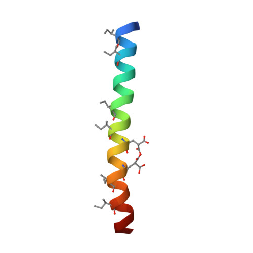
1TGG

RH3 DESIGNED RIGHT-HANDED COILED COIL TRIMER


Experimental Data Snapshot

  • Method: X-RAY DIFFRACTION
  • Resolution: 2.00 Å
  • R-Value Free: 0.269 
  • R-Value Work: 0.233 
  • R-Value Observed: 0.236 

wwPDB Validation   3D Report Full Report


This is version 1.2 of the entry. See complete history


Literature

Structural test of the parameterized-backbone method for protein design

Plecs, J.J.Harbury, P.B.Kim, P.S.Alber, T.

(2004) J Mol Biol 342: 289-297

  • DOI: https://doi.org/10.1016/j.jmb.2004.06.051
  • Primary Citation of Related Structures:  
    1TGG

  • PubMed Abstract: 

    Designing new protein folds requires a method for simultaneously optimizing the conformation of the backbone and the side-chains. One approach to this problem is the use of a parameterized backbone, which allows the systematic exploration of families of structures. We report the crystal structure of RH3, a right-handed, three-helix coiled coil that was designed using a parameterized backbone and detailed modeling of core packing. This crystal structure was determined using another rationally designed feature, a metal-binding site that permitted experimental phasing of the X-ray data. RH3 adopted the intended fold, which has not been observed previously in biological proteins. Unanticipated structural asymmetry in the trimer was a principal source of variation within the RH3 structure. The sequence of RH3 differs from that of a previously characterized right-handed tetramer, RH4, at only one position in each 11 amino acid sequence repeat. This close similarity indicates that the design method is sensitive to the core packing interactions that specify the protein structure. Comparison of the structures of RH3 and RH4 indicates that both steric overlap and cavity formation provide strong driving forces for oligomer specificity.


  • Organizational Affiliation

    Department of Physics, University of California, Berkeley, 94720, USA.


Macromolecules
Find similar proteins by:  (by identity cutoff)  |  3D Structure
Entity ID: 1
MoleculeChains Sequence LengthOrganismDetailsImage
right-handed coiled coil trimer
A, B, C
34N/AMutation(s): 0 
Entity Groups  
Sequence Clusters30% Identity50% Identity70% Identity90% Identity95% Identity100% Identity
Sequence Annotations
Expand
  • Reference Sequence
Small Molecules
Ligands 2 Unique
IDChains Name / Formula / InChI Key2D Diagram3D Interactions
NI
Query on NI

Download Ideal Coordinates CCD File 
D [auth B],
F [auth B],
I [auth B]
NICKEL (II) ION
Ni
VEQPNABPJHWNSG-UHFFFAOYSA-N
CL
Query on CL

Download Ideal Coordinates CCD File 
E [auth B],
G [auth B],
H [auth B],
J [auth B],
K [auth B]
CHLORIDE ION
Cl
VEXZGXHMUGYJMC-UHFFFAOYSA-M
Modified Residues  2 Unique
IDChains TypeFormula2D DiagramParent
CGU
Query on CGU
A, B, C
L-PEPTIDE LINKINGC6 H9 N O6GLU
IIL
Query on IIL
A, B, C
L-PEPTIDE LINKINGC6 H13 N O2ILE
Experimental Data & Validation

Experimental Data

  • Method: X-RAY DIFFRACTION
  • Resolution: 2.00 Å
  • R-Value Free: 0.269 
  • R-Value Work: 0.233 
  • R-Value Observed: 0.236 
  • Space Group: P 32
Unit Cell:
Length ( Å )Angle ( ˚ )
a = 25.42α = 90
b = 25.42β = 90
c = 141.34γ = 120
Software Package:
Software NamePurpose
MOSFLMdata reduction
SCALAdata scaling
MLPHAREphasing
X-PLORrefinement
CCP4data scaling

Structure Validation

View Full Validation Report


Entry History 

Deposition Data

Revision History  (Full details and data files)

  • Version 1.0: 2004-10-12
    Type: Initial release
  • Version 1.1: 2008-04-30
    Changes: Version format compliance
  • Version 1.2: 2011-07-13
    Changes: Version format compliance