1TEC | pdb_00001tec

CRYSTALLOGRAPHIC REFINEMENT BY INCORPORATION OF MOLECULAR DYNAMICS. THE THERMOSTABLE SERINE PROTEASE THERMITASE COMPLEXED WITH EGLIN-C


Experimental Data Snapshot

  • Method: X-RAY DIFFRACTION
  • Resolution: 2.20 Å
  • R-Value Work: 
    0.174 (DCC) 

wwPDB Validation 3D Report Full Report

Validation slider image for 1TEC

This is version 1.3 of the entry. See complete history

Literature

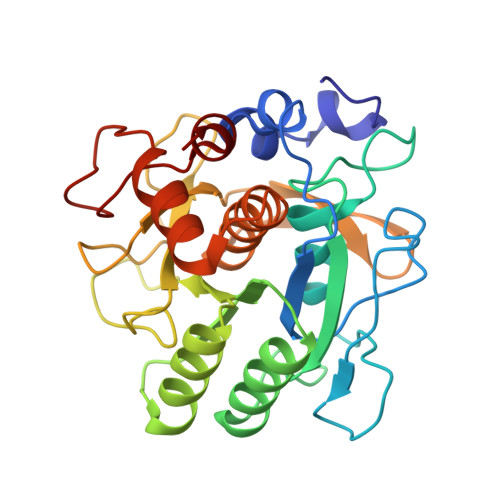
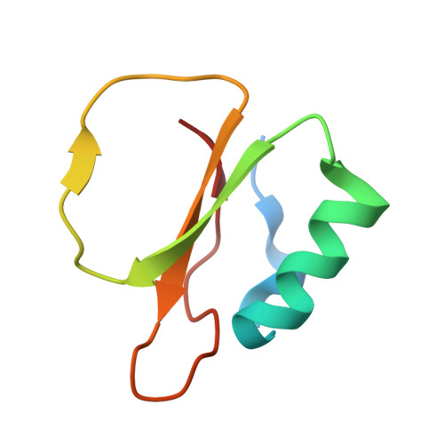
Crystallographic refinement by incorporation of molecular dynamics: thermostable serine protease thermitase complexed with eglin c.

Gros, P.Fujinaga, M.Dijkstra, B.W.Kalk, K.H.Hol, W.G.

(1989) Acta Crystallogr B 45: 488-499

  • DOI: https://doi.org/10.1107/s0108768189006038
  • Primary Citation Related Structures: 
    1TEC

  • PubMed Abstract: 

    In order to investigate the principles of protein thermostability, the crystal structure of thermitase from Thermoactinomyces vulgaris, a thermostable member of the subtilisin family of serine proteases, has been determined in a complex with eglin c. Eglin c is a serine protease inhibitor from the leech Hirudo medicinalis. After data collection with a television area-detector diffractometer and initial structure solution by molecular-replacement methods, crystallographic refinement proceeded with incorporation of molecular-dynamics techniques. It appeared that this refinement procedure has a large convergence radius with movements of more than 5 A for many atoms. Two procedures for the crystallographic molecular-dynamics refinement have been tested. They differed mainly in time span and weight on the X-ray 'energy'. The best results were obtained with a procedure which allowed the molecular-dynamics technique to search a large area in conformational space by having less weight on the X-ray restraints and allowing more time. The use of molecular-dynamics refinement considerably simplified the laborious and difficult task of fitting the model in its electron density during the refinement process. The final crystallographic R factor is 17.9% at 2.2 A resolution.


  • Organizational Affiliation
    • Department of Chemistry, University of Groningen, The Netherlands.

Macromolecule Content 

  • Total Structure Weight: 36.59 kDa 
  • Atom Count: 2,737 
  • Modeled Residue Count: 342 
  • Deposited Residue Count: 349 
  • Unique protein chains: 2

Macromolecules

Find similar proteins by:|  3D Structure
Entity ID: 1
MoleculeChains  Sequence LengthOrganismDetailsImage
THERMITASEA [auth E]279Thermoactinomyces vulgarisMutation(s): 0 
EC: 3.4.21.66
UniProt
Find proteins for P04072 (Thermoactinomyces vulgaris)
Explore P04072 
Go to UniProtKB:  P04072
Entity Groups
Sequence Clusters30% Identity50% Identity70% Identity90% Identity95% Identity100% Identity
UniProt GroupP04072
Sequence Annotations
Expand
Reference Sequence
Find similar proteins by:|  3D Structure
Entity ID: 2
MoleculeChains  Sequence LengthOrganismDetailsImage
EGLIN CB [auth I]70Hirudo medicinalisMutation(s): 0 
UniProt
Find proteins for P01051 (Hirudo medicinalis)
Explore P01051 
Go to UniProtKB:  P01051
Entity Groups
Sequence Clusters30% Identity50% Identity70% Identity90% Identity95% Identity100% Identity
UniProt GroupP01051
Sequence Annotations
Expand
Reference Sequence

Experimental Data & Validation

Experimental Data

  • Method: X-RAY DIFFRACTION
  • Resolution: 2.20 Å
  • R-Value Work:  0.174 (DCC) 
Space Group: P 21 21 21
Unit Cell:
Length ( Å )Angle ( ˚ )
a = 63.25α = 90
b = 72.1β = 90
c = 89.25γ = 90
Software Package:
Software NamePurpose
GROMOSrefinement

Structure Validation

View Full Validation Report


Entry History 

Deposition Data

Revision History  (Full details and data files)

  • Version 1.0: 1989-10-15
    Type: Initial release
  • Version 1.1: 2008-03-24
    Changes: Version format compliance
  • Version 1.2: 2011-07-13
    Changes: Version format compliance
  • Version 1.3: 2024-02-14
    Changes: Data collection, Database references, Derived calculations, Other