1TDT | pdb_00001tdt

THREE-DIMENSIONAL STRUCTURE OF TETRAHYDRODIPICOLINATE-N-SUCCINYLTRANSFERASE


Experimental Data Snapshot

  • Method: X-RAY DIFFRACTION
  • Resolution: 2.20 Å
  • R-Value Free: 
    0.230 (Depositor) 
  • R-Value Work: 
    0.170 (Depositor), 0.160 (DCC) 

wwPDB Validation 3D Report Full Report

Validation slider image for 1TDT

This is version 1.3 of the entry. See complete history

Literature

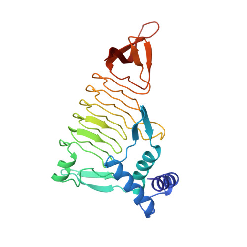
Three-dimensional structure of tetrahydrodipicolinate N-succinyltransferase.

Beaman, T.W.Binder, D.A.Blanchard, J.S.Roderick, S.L.

(1997) Biochemistry 36: 489-494

  • DOI: https://doi.org/10.1021/bi962522q
  • Primary Citation Related Structures: 
    1TDT

  • PubMed Abstract: 

    The conversion of tetrahydrodipicolinate and succinyl-CoA to N-succinyltetrahydrodipicolinate and CoA is catalyzed by tetrahydrodipicolinate N-succinyltransferase and is the committed step in the succinylase pathway by which bacteria synthesize L-lysine and meso-diaminopimelate, a component of peptidoglycan. The X-ray crystal structure of THDP succinyltransferase has been determined to 2.2 A resolution and has been refined to a crystallographic R-factor of 17.0%. The enzyme is trimeric and displays the left-handed parallel beta-helix (L beta H) structural motif encoded by the "hexapeptide repeat" amino acid sequence motif [Raetz, C.R.H., & Roderick, S.L. (1995) Science 270, 997-1000]. The approximate location of the active site of THDP succinyltransferase is suggested by the proximity of binding sites for two inhibitors: p-(chloromercuri)benzenesulfonic acid and cobalt ion, both of which bind to the L beta H domain.


  • Organizational Affiliation
    • Department of Biochemistry, Albert Einstein College of Medicine, Bronx, New York 10461, USA.

Macromolecule Content 

  • Total Structure Weight: 84.75 kDa 
  • Atom Count: 5,995 
  • Modeled Residue Count: 764 
  • Deposited Residue Count: 777 
  • Unique protein chains: 1

Macromolecules

Find similar proteins by:|  3D Structure
Entity ID: 1
MoleculeChains  Sequence LengthOrganismDetailsImage
TETRAHYDRODIPICOLINATE-N-SUCCINYLTRANSFERASE
A, B, C
259Mycobacterium tuberculosis variant bovisMutation(s): 0 
EC: 2.3.1.117
UniProt
Find proteins for P56220 (Unknown prokaryotic organism)
Explore P56220 
Go to UniProtKB:  P56220
Entity Groups
Sequence Clusters30% Identity50% Identity70% Identity90% Identity95% Identity100% Identity
UniProt GroupP56220
Sequence Annotations
Expand
Reference Sequence

Experimental Data & Validation

Experimental Data

  • Method: X-RAY DIFFRACTION
  • Resolution: 2.20 Å
  • R-Value Free:  0.230 (Depositor) 
  • R-Value Work:  0.170 (Depositor), 0.160 (DCC) 
Space Group: P 1 21 1
Unit Cell:
Length ( Å )Angle ( ˚ )
a = 60.6α = 90
b = 107.9β = 113
c = 70.7γ = 90
Software Package:
Software NamePurpose
PHASESphasing
TNTrefinement
XDSdata reduction
XSCALEdata scaling

Structure Validation

View Full Validation Report


Entry History 

Deposition Data

Revision History  (Full details and data files)

  • Version 1.0: 1997-06-05
    Type: Initial release
  • Version 1.1: 2008-03-24
    Changes: Version format compliance
  • Version 1.2: 2011-07-13
    Changes: Version format compliance
  • Version 1.3: 2024-02-14
    Changes: Data collection, Database references