1TD0 | pdb_00001td0

Viral capsid protein SHP at pH 5.5


Experimental Data Snapshot

  • Method: X-RAY DIFFRACTION
  • Resolution: 1.95 Å
  • R-Value Free: 
    0.260 (Depositor), 0.260 (DCC) 
  • R-Value Work: 
    0.229 (Depositor), 0.230 (DCC) 
  • R-Value Observed: 
    0.229 (Depositor) 

wwPDB Validation 3D Report Full Report

Validation slider image for 1TD0

This is version 1.3 of the entry. See complete history

Literature

Kinetic Stability and Crystal Structure of the Viral Capsid Protein SHP

Forrer, P.Chang, C.Ott, D.Wlodawer, A.Plueckthun, A.

(2004) J Mol Biology 344: 179-193

  • DOI: https://doi.org/10.1016/j.jmb.2004.09.030
  • Primary Citation Related Structures: 
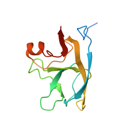
    1TD0, 1TD3, 1TD4

  • PubMed Abstract: 

    SHP, the capsid-stabilizing protein of lambdoid phage 21, is highly resistant against denaturant-induced unfolding. We demonstrate that this high functional stability of SHP is due to a high kinetic stability with a half-life for unfolding of 25 days at zero denaturant, while the thermodynamic stability is not unusually high. Unfolding experiments demonstrated that the trimeric state (also observed in crystals and present on the phage capsid) of SHP is kinetically stable in solution, while the monomer intermediate unfolds very rapidly. We also determined the crystal structure of trimeric SHP at 1.5A resolution, which was compared to that of its functional homolog gpD. This explains how a tight network of H-bonds rigidifies crucial interpenetrating residues, leading to the observed extremely slow trimer dissociation or denaturation. Taken as a whole, our results provide molecular-level insights into natural strategies to achieve kinetic stability by taking advantage of protein oligomerization. Kinetic stability may be especially needed in phage capsids to allow survival in harsh environments.


  • Organizational Affiliation
    • Biochemisches Institut, Universität Zürich, Winterthurerstrasse 190, CH-8057 Zürich, Switzerland.

Macromolecule Content 

  • Total Structure Weight: 47.43 kDa 
  • Atom Count: 3,536 
  • Modeled Residue Count: 408 
  • Deposited Residue Count: 456 
  • Unique protein chains: 1

Macromolecules

Find similar proteins by:|  3D Structure
Entity ID: 1
MoleculeChains  Sequence LengthOrganismDetailsImage
Head decoration protein
A, B, C, D
114Enterobacteria phage P21Mutation(s): 0 
UniProt
Find proteins for P36275 (Enterobacteria phage P21)
Explore P36275 
Go to UniProtKB:  P36275
Entity Groups
Sequence Clusters30% Identity50% Identity70% Identity90% Identity95% Identity100% Identity
UniProt GroupP36275
Sequence Annotations
Expand
Reference Sequence

Experimental Data & Validation

Experimental Data

  • Method: X-RAY DIFFRACTION
  • Resolution: 1.95 Å
  • R-Value Free:  0.260 (Depositor), 0.260 (DCC) 
  • R-Value Work:  0.229 (Depositor), 0.230 (DCC) 
  • R-Value Observed: 0.229 (Depositor) 
Space Group: P 3
Unit Cell:
Length ( Å )Angle ( ˚ )
a = 57.482α = 90
b = 57.482β = 90
c = 101.608γ = 120
Software Package:
Software NamePurpose
CNSrefinement
DENZOdata reduction
SCALEPACKdata scaling
AMoREphasing

Structure Validation

View Full Validation Report


Entry History 

Deposition Data

Revision History  (Full details and data files)

  • Version 1.0: 2004-11-02
    Type: Initial release
  • Version 1.1: 2008-04-30
    Changes: Version format compliance
  • Version 1.2: 2011-07-13
    Changes: Version format compliance
  • Version 1.3: 2024-02-14
    Changes: Data collection, Database references