1TC8

Crystal structure of Krait-venom phospholipase A2 in a complex with a natural fatty acid tridecanoic acid


Experimental Data Snapshot

  • Method: X-RAY DIFFRACTION
  • Resolution: 2.70 Å
  • R-Value Free: 0.233 
  • R-Value Work: 0.204 
  • R-Value Observed: 0.204 

wwPDB Validation   3D Report Full Report


This is version 1.3 of the entry. See complete history


Literature

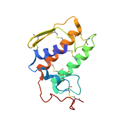
Crystal structure of the complex formed between a group I phospholipase A2 and a naturally occurring fatty acid at 2.7 A resolution

Singh, G.Jasti, J.Saravanan, K.Sharma, S.Kaur, P.Srinivasan, A.Singh, T.P.

(2005) Protein Sci 14: 395-400

  • DOI: https://doi.org/10.1110/ps.041115505
  • Primary Citation of Related Structures:  
    1TC8

  • PubMed Abstract: 

    This is the first evidence of a naturally bound fatty acid to a group I Phospholipase A(2) (PLA(2)) and also to a PLA(2) with Asp 49. The fatty acid identified as n-tridecanoic acid is observed at the substrate recognition site of PLA(2) hydrophobic channel. The complex was isolated from the venom of Bungarus caeruleus (Common Indian Krait). The primary sequence of the PLA(2) was determined using the cDNA method. Three-dimensional structure has been solved by the molecular replacement method and refined using the CNS package to a final R factor of 19.8% for the data in the resolution range from 20.0 to 2.7 A. The final refined model is comprised of 912 protein atoms, one sodium ion, one molecule of n-tridecanoic acid, and 60 water molecules. The sodium ion is located in the calcium-binding loop with a sevenfold coordination. A characteristic extra electron density was observed in the hydrophobic channel of the enzyme, into which a molecule of n-tridecanoic acid was clearly fitted. The MALDI-TOF measurements of the crystals had earlier indicated an increase in the molecular mass of PLA(2) by 212 Da over the native PLA(2). A major part of the ligand fits well in the binding pocket and interacts directly with His 48 and Asp 49. Although the overall structure of PLA(2) in the present complex is similar to the native structure reported earlier, it differs significantly in the folding of its calcium-binding loop.


  • Organizational Affiliation

    Department of Biophysics, All India Institute of Medical Sciences, Ansari Nagar, New Delhi, 110029, India. tps@aiims.aiims.ac.in.


Macromolecules
Find similar proteins by:  (by identity cutoff)  |  3D Structure
Entity ID: 1
MoleculeChains Sequence LengthOrganismDetailsImage
phospholipase A2 isoform 1118Bungarus caeruleusMutation(s): 0 
EC: 3.1.1.4
Membrane Entity: Yes 
UniProt
Find proteins for Q6SLM2 (Bungarus caeruleus)
Explore Q6SLM2 
Go to UniProtKB:  Q6SLM2
Entity Groups  
Sequence Clusters30% Identity50% Identity70% Identity90% Identity95% Identity100% Identity
UniProt GroupQ6SLM2
Sequence Annotations
Expand
  • Reference Sequence
Small Molecules
Ligands 2 Unique
IDChains Name / Formula / InChI Key2D Diagram3D Interactions
TDA
Query on TDA

Download Ideal Coordinates CCD File 
C [auth A]N-TRIDECANOIC ACID
C13 H26 O2
SZHOJFHSIKHZHA-UHFFFAOYSA-N
NA
Query on NA

Download Ideal Coordinates CCD File 
B [auth A]SODIUM ION
Na
FKNQFGJONOIPTF-UHFFFAOYSA-N
Experimental Data & Validation

Experimental Data

  • Method: X-RAY DIFFRACTION
  • Resolution: 2.70 Å
  • R-Value Free: 0.233 
  • R-Value Work: 0.204 
  • R-Value Observed: 0.204 
  • Space Group: P 41 21 2
Unit Cell:
Length ( Å )Angle ( ˚ )
a = 53.79α = 90
b = 53.79β = 90
c = 82.5γ = 90
Software Package:
Software NamePurpose
CNSrefinement
DENZOdata reduction
SCALEPACKdata scaling
AMoREphasing

Structure Validation

View Full Validation Report


Entry History 

Deposition Data

Revision History  (Full details and data files)

  • Version 1.0: 2004-06-08
    Type: Initial release
  • Version 1.1: 2008-04-30
    Changes: Version format compliance
  • Version 1.2: 2011-07-13
    Changes: Version format compliance
  • Version 1.3: 2023-10-25
    Changes: Data collection, Database references, Derived calculations, Refinement description