1TBG

BETA-GAMMA DIMER OF THE HETEROTRIMERIC G-PROTEIN TRANSDUCIN


Experimental Data Snapshot

  • Method: X-RAY DIFFRACTION
  • Resolution: 2.10 Å
  • R-Value Free: 0.306 
  • R-Value Work: 0.200 
  • R-Value Observed: 0.200 

wwPDB Validation   3D Report Full Report


This is version 1.3 of the entry. See complete history


Literature

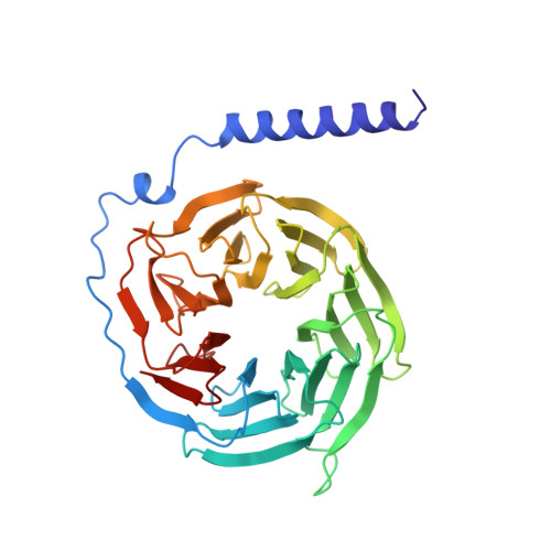
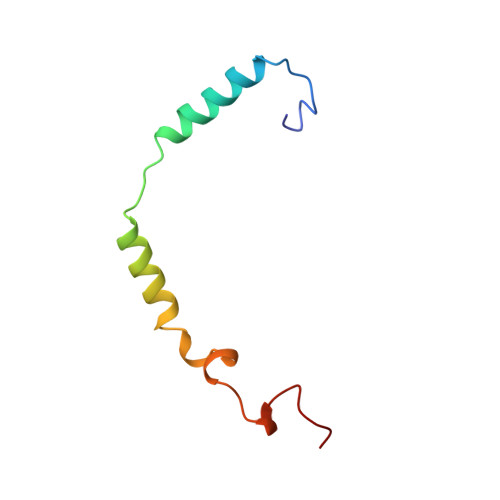
Crystal structure of a G-protein beta gamma dimer at 2.1A resolution.

Sondek, J.Bohm, A.Lambright, D.G.Hamm, H.E.Sigler, P.B.

(1996) Nature 379: 369-374

  • DOI: https://doi.org/10.1038/379369a0
  • Primary Citation of Related Structures:  
    1TBG

  • PubMed Abstract: 

    Many signalling cascades use seven-helical transmembrane receptors coupled to heterotrimeric G proteins (G alpha beta gamma) to convert extracellular signals into intracellular responses. Upon nucleotide exchange catalysed by activated receptors, heterotrimers dissociate into GTP-bound G alpha subunits and G beta gamma dimers, either of which can modulate many downstream effectors. Here we use multiwavelength anomalous diffraction data to solve the crystal structure of the beta gamma dimer of the G protein transducin. The beta-subunit is primarily a seven-bladed beta-propeller that is partially encircled by an extended gamma-subunit. The beta-propeller, which contains seven structurally similar WD repeats, defines the stereochemistry of the WD repeat and the probable architecture of all WD-repeat-containing domains. The structure details interactions between G protein beta- and gamma-subunits and highlights regions implicated in effector modulation for the conserved family of G protein beta gamma dimers.


  • Organizational Affiliation

    Department of Molecular Biophysics and Biochemistry, Yale University, New Haven, Connecticut 06510, USA.


Macromolecules
Find similar proteins by:  (by identity cutoff)  |  3D Structure
Entity ID: 1
MoleculeChains Sequence LengthOrganismDetailsImage
TRANSDUCINA,
C [auth B],
E [auth C],
G [auth D]
340Bos taurusMutation(s): 0 
UniProt
Find proteins for P62871 (Bos taurus)
Explore P62871 
Go to UniProtKB:  P62871
Entity Groups  
Sequence Clusters30% Identity50% Identity70% Identity90% Identity95% Identity100% Identity
UniProt GroupP62871
Sequence Annotations
Expand
  • Reference Sequence
Find similar proteins by:  (by identity cutoff)  |  3D Structure
Entity ID: 2
MoleculeChains Sequence LengthOrganismDetailsImage
TRANSDUCINB [auth E],
D [auth F],
F [auth G],
H
68Bos taurusMutation(s): 0 
UniProt
Find proteins for P02698 (Bos taurus)
Explore P02698 
Go to UniProtKB:  P02698
Entity Groups  
Sequence Clusters30% Identity50% Identity70% Identity90% Identity95% Identity100% Identity
UniProt GroupP02698
Sequence Annotations
Expand
  • Reference Sequence
Experimental Data & Validation

Experimental Data

  • Method: X-RAY DIFFRACTION
  • Resolution: 2.10 Å
  • R-Value Free: 0.306 
  • R-Value Work: 0.200 
  • R-Value Observed: 0.200 
  • Space Group: P 21 21 21
Unit Cell:
Length ( Å )Angle ( ˚ )
a = 85.1α = 90
b = 94β = 90
c = 194.7γ = 90
Software Package:
Software NamePurpose
X-PLORmodel building
X-PLORrefinement
DENZOdata reduction
SCALEPACKdata scaling
X-PLORphasing

Structure Validation

View Full Validation Report


Entry History 

Deposition Data

Revision History  (Full details and data files)

  • Version 1.0: 1997-04-01
    Type: Initial release
  • Version 1.1: 2008-03-24
    Changes: Version format compliance
  • Version 1.2: 2011-07-13
    Changes: Derived calculations, Version format compliance
  • Version 1.3: 2024-02-14
    Changes: Data collection, Database references, Other