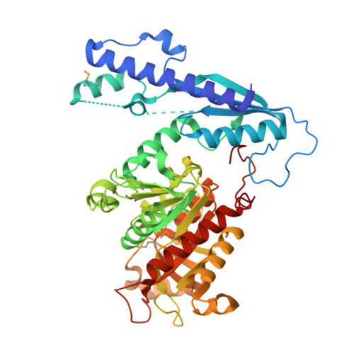
1T8S

Crystal Structure of E.coli AMP Nucleosidase complexed with formicin 5'-monophosphate


Experimental Data Snapshot

  • Method: X-RAY DIFFRACTION
  • Resolution: 2.60 Å
  • R-Value Free: 0.256 
  • R-Value Work: 0.226 
  • R-Value Observed: 0.226 

wwPDB Validation   3D Report Full Report


This is version 1.3 of the entry. See complete history


Literature

Structure of Escherichia coli AMP Nucleosidase Reveals Similarity to Nucleoside Phosphorylases

Zhang, Y.Cottet, S.E.Ealick, S.E.

(2004) Structure 12: 1383-1394

  • DOI: https://doi.org/10.1016/j.str.2004.05.015
  • Primary Citation of Related Structures:  
    1T8R, 1T8S, 1T8W, 1T8Y

  • PubMed Abstract: 

    AMP nucleosidase (AMN) catalyzes the hydrolysis of AMP to form adenine and ribose 5-phosphate. The enzyme is found only in prokaryotes, where it plays a role in purine nucleoside salvage and intracellular AMP level regulation. Enzyme activity is stimulated by ATP and suppressed by phosphate. The structure of unliganded AMN was determined at 2.7 A resolution, and structures of the complexes with either formycin 5'-monophosphate or inorganic phosphate were determined at 2.6 A and 3.0 A resolution, respectively. AMN is a biological homohexamer, and each monomer is composed of two domains: a catalytic domain and a putative regulatory domain. The overall topology of the catalytic domain and some features of the substrate binding site resemble those of the nucleoside phosphorylases, demonstrating that AMN is a new member of the family. The structure of the regulatory domain consists of a long helix and a four-stranded sheet and has a novel topology.


  • Organizational Affiliation

    Department of Chemistry and Chemical Biology, Cornell University, Ithaca, New York 14853, USA.


Macromolecules
Find similar proteins by:  (by identity cutoff)  |  3D Structure
Entity ID: 1
MoleculeChains Sequence LengthOrganismDetailsImage
AMP nucleosidase
A, B, C, D, E
A, B, C, D, E, F
484Escherichia coliMutation(s): 4 
Gene Names: AMNB1982Z3139ECS2779
EC: 3.2.2.4
UniProt
Find proteins for P0AE12 (Escherichia coli (strain K12))
Explore P0AE12 
Go to UniProtKB:  P0AE12
Entity Groups  
Sequence Clusters30% Identity50% Identity70% Identity90% Identity95% Identity100% Identity
UniProt GroupP0AE12
Sequence Annotations
Expand
  • Reference Sequence
Experimental Data & Validation

Experimental Data

  • Method: X-RAY DIFFRACTION
  • Resolution: 2.60 Å
  • R-Value Free: 0.256 
  • R-Value Work: 0.226 
  • R-Value Observed: 0.226 
  • Space Group: P 42 21 2
Unit Cell:
Length ( Å )Angle ( ˚ )
a = 246.24α = 90
b = 246.24β = 90
c = 111.13γ = 90
Software Package:
Software NamePurpose
CNSrefinement
HKL-2000data reduction
SCALEPACKdata scaling
CNSphasing

Structure Validation

View Full Validation Report


Entry History 

Deposition Data

Revision History  (Full details and data files)

  • Version 1.0: 2004-08-17
    Type: Initial release
  • Version 1.1: 2008-04-30
    Changes: Version format compliance
  • Version 1.2: 2011-07-13
    Changes: Version format compliance
  • Version 1.3: 2024-10-16
    Changes: Data collection, Database references, Derived calculations, Structure summary