


The crystal structure of Glycerophosphoryl diester phosphodiesterase from E. coli
Zhang, R., Kim, Y., Dementieva, I., Duke, N., Stols, L., Donnelly, M., Joachimiak, A.To be published.
Experimental Data Snapshot
wwPDB Validation 3D Report Full Report

Entity ID: 1 | |||||
|---|---|---|---|---|---|
| Molecule | Chains | Sequence Length | Organism | Details | Image |
| Glycerophosphoryl diester phosphodiesterase, periplasmic | 336 | Escherichia coli | Mutation(s): 9 Gene Names: GLPQ, B2239 EC: 3.1.4.46 | 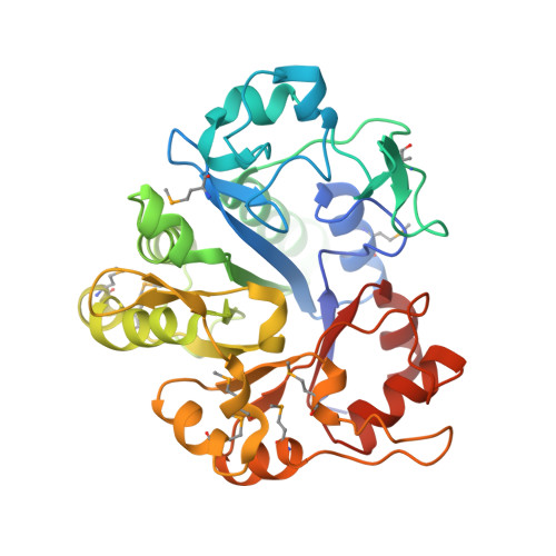 | |
UniProt | |||||
Entity Groups | |||||
| Sequence Clusters | 30% Identity50% Identity70% Identity90% Identity95% Identity100% Identity | ||||
| UniProt Group | P09394 | ||||
Sequence AnnotationsExpand | |||||
Reference Sequence | |||||
| Ligands 2 Unique | |||||
|---|---|---|---|---|---|
| ID | Chains | Name / Formula / InChI Key | 2D Diagram | 3D Interactions | |
| GOL Download:Ideal Coordinates CCD File | F [auth A] G [auth A] H [auth A] J [auth B] K [auth B] | GLYCEROL C3 H8 O3 PEDCQBHIVMGVHV-UHFFFAOYSA-N |  | ||
| MG Download:Ideal Coordinates CCD File | E [auth A], I [auth B], L [auth C], P [auth D] | MAGNESIUM ION Mg JLVVSXFLKOJNIY-UHFFFAOYSA-N |  | ||
| Modified Residues 1 Unique | |||||
|---|---|---|---|---|---|
| ID | Chains | Type | Formula | 2D Diagram | Parent |
| MSE Query on MSE | A, B, C, D | L-PEPTIDE LINKING | C5 H11 N O2 Se |  | MET |
| Length ( Å ) | Angle ( ˚ ) |
|---|---|
| a = 62.759 | α = 90 |
| b = 118.386 | β = 90 |
| c = 242.048 | γ = 90 |
| Software Name | Purpose |
|---|---|
| CNS | refinement |
| SBC-Collect | data collection |
| HKL-2000 | data scaling |
| CNS | phasing |


