1T3Q

Crystal structure of quinoline 2-Oxidoreductase from Pseudomonas Putida 86


Experimental Data Snapshot

  • Method: X-RAY DIFFRACTION
  • Resolution: 1.80 Å
  • R-Value Free: 0.207 
  • R-Value Work: 0.186 
  • R-Value Observed: 0.186 

Starting Model: experimental
View more details

wwPDB Validation   3D Report Full Report


Ligand Structure Quality Assessment 


This is version 1.4 of the entry. See complete history


Literature

Active site geometry and substrate recognition of the molybdenum hydroxylase quinoline 2-oxidoreductase.

Bonin, I.Martins, B.M.Purvanov, V.Fetzner, S.Huber, R.Dobbek, H.

(2004) Structure 12: 1425-1435

  • DOI: https://doi.org/10.1016/j.str.2004.05.014
  • Primary Citation of Related Structures:  
    1T3Q

  • PubMed Abstract: 

    The soil bacterium Pseudomonas putida 86 uses quinoline as a sole source of carbon and energy. Quinoline 2-oxidoreductase (Qor) catalyzes the first metabolic step converting quinoline to 2-oxo-1,2-dihydroquinoline. Qor is a member of the molybdenum hydroxylases. The molybdenum ion is coordinated by two ene-dithiolate sulfur atoms, two oxo-ligands, and a catalytically crucial sulfido-ligand, whose position in the active site was controversial. The 1.8 A resolution crystal structure of Qor indicates that the sulfido-ligand occupies the equatorial position at the molybdenum ion. The structural comparison of Qor with the allopurinol-inhibited xanthine dehydrogenase from Rhodobacter capsulatus allows direct insight into the mechanism of substrate recognition and the identification of putative catalytic residues. The active site protein variants QorE743V and QorE743D were analyzed to assess the catalytic role of E743.


  • Organizational Affiliation

    Abteilung für Strukturforschung, Max-Planck-Institut für Biochemie, 82152 Martinsried, Germany.


Macromolecules
Find similar proteins by:  (by identity cutoff)  |  3D Structure
Entity ID: 1
MoleculeChains Sequence LengthOrganismDetailsImage
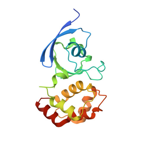
quinoline 2-oxidoreductase small subunit
A, D
168Pseudomonas putidaMutation(s): 0 
EC: 1.3.99.17
UniProt
Find proteins for P72223 (Pseudomonas putida)
Explore P72223 
Go to UniProtKB:  P72223
Entity Groups  
Sequence Clusters30% Identity50% Identity70% Identity90% Identity95% Identity100% Identity
UniProt GroupP72223
Sequence Annotations
Expand
  • Reference Sequence
Find similar proteins by:  (by identity cutoff)  |  3D Structure
Entity ID: 2
MoleculeChains Sequence LengthOrganismDetailsImage
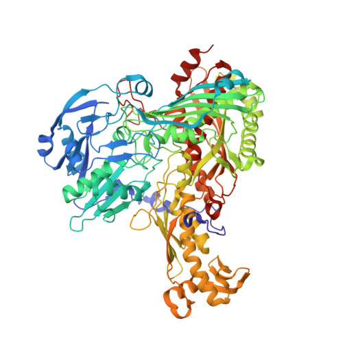
quinoline 2-oxidoreductase large subunit
B, E
788Pseudomonas putidaMutation(s): 0 
EC: 1.3.99.17
UniProt
Find proteins for P72224 (Pseudomonas putida)
Explore P72224 
Go to UniProtKB:  P72224
Entity Groups  
Sequence Clusters30% Identity50% Identity70% Identity90% Identity95% Identity100% Identity
UniProt GroupP72224
Sequence Annotations
Expand
  • Reference Sequence
Find similar proteins by:  (by identity cutoff)  |  3D Structure
Entity ID: 3
MoleculeChains Sequence LengthOrganismDetailsImage
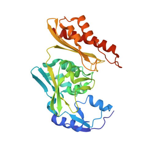
quinoline 2-oxidoreductase medium subunit
C, F
288Pseudomonas putidaMutation(s): 0 
EC: 1.3.99.17
UniProt
Find proteins for P72222 (Pseudomonas putida)
Explore P72222 
Go to UniProtKB:  P72222
Entity Groups  
Sequence Clusters30% Identity50% Identity70% Identity90% Identity95% Identity100% Identity
UniProt GroupP72222
Sequence Annotations
Expand
  • Reference Sequence
Small Molecules
Ligands 6 Unique
IDChains Name / Formula / InChI Key2D Diagram3D Interactions
FAD
Query on FAD

Download Ideal Coordinates CCD File 
FA [auth F],
R [auth C]
FLAVIN-ADENINE DINUCLEOTIDE
C27 H33 N9 O15 P2
VWWQXMAJTJZDQX-UYBVJOGSSA-N
MCN
Query on MCN

Download Ideal Coordinates CCD File 
AA [auth E],
N [auth B]
PTERIN CYTOSINE DINUCLEOTIDE
C19 H22 N8 O13 P2 S2
RBWYFPNWTRZKKZ-LOIMWUFNSA-N
FES
Query on FES

Download Ideal Coordinates CCD File 
G [auth A],
H [auth A],
U [auth D],
V [auth D]
FE2/S2 (INORGANIC) CLUSTER
Fe2 S2
NIXDOXVAJZFRNF-UHFFFAOYSA-N
SMO
Query on SMO

Download Ideal Coordinates CCD File 
BA [auth E],
O [auth B]
DIOXOSULFIDOMOLYBDENUM(VI) ION
Mo O2 S
BSDYLDOYZVCHEU-UHFFFAOYSA-N
SO4
Query on SO4

Download Ideal Coordinates CCD File 
J [auth B]
K [auth B]
L [auth B]
M [auth B]
W [auth E]
J [auth B],
K [auth B],
L [auth B],
M [auth B],
W [auth E],
X [auth E],
Y [auth E],
Z [auth E]
SULFATE ION
O4 S
QAOWNCQODCNURD-UHFFFAOYSA-L
GOL
Query on GOL

Download Ideal Coordinates CCD File 
CA [auth E]
DA [auth E]
EA [auth E]
GA [auth F]
I [auth A]
CA [auth E],
DA [auth E],
EA [auth E],
GA [auth F],
I [auth A],
P [auth B],
Q [auth B],
S [auth C],
T [auth C]
GLYCEROL
C3 H8 O3
PEDCQBHIVMGVHV-UHFFFAOYSA-N
Experimental Data & Validation

Experimental Data

  • Method: X-RAY DIFFRACTION
  • Resolution: 1.80 Å
  • R-Value Free: 0.207 
  • R-Value Work: 0.186 
  • R-Value Observed: 0.186 
  • Space Group: C 1 2 1
Unit Cell:
Length ( Å )Angle ( ˚ )
a = 278.32α = 90
b = 72.1β = 127.98
c = 202.65γ = 90
Software Package:
Software NamePurpose
MAR345data collection
XDSdata reduction
AMoREphasing
CNSrefinement
XDSdata scaling

Structure Validation

View Full Validation Report


Ligand Structure Quality Assessment 


Entry History 

Deposition Data

Revision History  (Full details and data files)

  • Version 1.0: 2004-09-14
    Type: Initial release
  • Version 1.1: 2008-04-30
    Changes: Version format compliance
  • Version 1.2: 2011-07-13
    Changes: Non-polymer description, Version format compliance
  • Version 1.3: 2017-10-11
    Changes: Advisory, Refinement description
  • Version 1.4: 2023-08-23
    Changes: Advisory, Data collection, Database references, Derived calculations, Refinement description