1T27 | pdb_00001t27

THE STRUCTURE OF PITP COMPLEXED TO PHOSPHATIDYLCHOLINE


Experimental Data Snapshot

  • Method: X-RAY DIFFRACTION
  • Resolution: 2.20 Å
  • R-Value Free: 
    0.250 (Depositor), 0.251 (DCC) 
  • R-Value Work: 
    0.205 (Depositor), 0.202 (DCC) 
  • R-Value Observed: 
    0.205 (Depositor) 

wwPDB Validation 3D Report Full Report

Validation slider image for 1T27

Ligand Structure Quality Assessment 


This is version 1.3 of the entry. See complete history

Literature

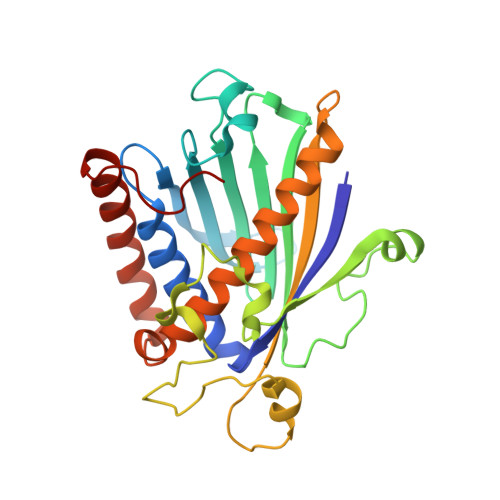
STRUCTURE OF A MULTIFUNCTIONAL PROTEIN. MAMMALIAN PHOSPHATIDYLINOSITOL TRANSFER PROTEIN COMPLEXED WITH PHOSPHATIDYLCHOLINE

Yoder, M.D.Thomas, L.M.Tremblay, J.M.Oliver, R.L.Yarbrough, L.R.Helmkamp Jr., G.M.

(2001) J Biological Chem 276: 9246-9252

  • DOI: https://doi.org/10.1074/jbc.M010131200
  • Primary Citation Related Structures: 
    1T27

  • PubMed Abstract: 

    Eukaryotic phosphatidylinositol transfer protein is a ubiquitous multifunctional protein that transports phospholipids between membrane surfaces and participates in cellular phospholipid metabolism during signal transduction and vesicular trafficking. The three-dimensional structure of the alpha-isoform of rat phosphatidylinositol transfer protein complexed with one molecule of phosphatidylcholine, one of its physiological ligands, has been determined to 2.2 A resolution by x-ray diffraction techniques. A single beta-sheet and several long alpha-helices define an enclosed internal cavity in which a single molecule of the phospholipid is accommodated with its polar head group in the center of the protein and fatty acyl chains projected toward the surface. Other structural features suggest mechanisms by which cytosolic phosphatidylinositol transfer protein interacts with membranes for lipid exchange and associates with a variety of lipid and protein kinases.


  • Organizational Affiliation
    • Division of Cell Biology and Biophysics, School of Biological Sciences, University of Missouri-Kansas City, Kansas City, Missouri 64110-2499 , USA. yoderm@umkc.edu

Macromolecule Content 

  • Total Structure Weight: 32.74 kDa 
  • Atom Count: 2,429 
  • Modeled Residue Count: 269 
  • Deposited Residue Count: 271 
  • Unique protein chains: 1

Macromolecules

Find similar proteins by:|  3D Structure
Entity ID: 1
MoleculeChains  Sequence LengthOrganismDetailsImage
Phosphatidylinositol transfer protein alpha isoform271Rattus norvegicusMutation(s): 0 
Gene Names: PITPNPITPNA
UniProt
Find proteins for P16446 (Rattus norvegicus)
Explore P16446 
Go to UniProtKB:  P16446
Entity Groups
Sequence Clusters30% Identity50% Identity70% Identity90% Identity95% Identity100% Identity
UniProt GroupP16446
Sequence Annotations
Expand
Reference Sequence

Small Molecules

Ligands 1 Unique
IDChains Name / Formula / InChI Key2D Diagram3D Interactions
PCW

Query on PCW


Download:Ideal Coordinates CCD File
B [auth A]1,2-DIOLEOYL-SN-GLYCERO-3-PHOSPHOCHOLINE
C44 H85 N O8 P
SNKAWJBJQDLSFF-NVKMUCNASA-O

Experimental Data & Validation

Experimental Data

  • Method: X-RAY DIFFRACTION
  • Resolution: 2.20 Å
  • R-Value Free:  0.250 (Depositor), 0.251 (DCC) 
  • R-Value Work:  0.205 (Depositor), 0.202 (DCC) 
  • R-Value Observed: 0.205 (Depositor) 
Space Group: P 1 21 1
Unit Cell:
Length ( Å )Angle ( ˚ )
a = 43.914α = 90
b = 73.773β = 114.73
c = 48.185γ = 90
Software Package:
Software NamePurpose
CNSrefinement
DENZOdata reduction
SCALEPACKdata scaling
SHARPphasing

Structure Validation

View Full Validation Report


Ligand Structure Quality Assessment 


Entry History 

Deposition Data

Revision History  (Full details and data files)

  • Version 1.0: 2004-05-11
    Type: Initial release
  • Version 1.1: 2008-04-30
    Changes: Version format compliance
  • Version 1.2: 2011-07-13
    Changes: Version format compliance
  • Version 1.3: 2024-02-14
    Changes: Data collection, Database references, Derived calculations