1SZH | pdb_00001szh

Crystal Structure of C. elegans HER-1


Experimental Data Snapshot

  • Method: X-RAY DIFFRACTION
  • Resolution: 1.50 Å
  • R-Value Free: 
    0.227 (Depositor), 0.227 (DCC) 
  • R-Value Work: 
    0.209 (Depositor), 0.208 (DCC) 
  • R-Value Observed: 
    0.209 (Depositor) 

wwPDB Validation 3D Report Full Report

Validation slider image for 1SZH

This is version 1.4 of the entry. See complete history

Literature

Crystal structure of Caenorhabditis elegans HER-1 and characterization of the interaction between HER-1 and TRA-2A.

Hamaoka, B.Y.Dann III, C.E.Geisbrecht, B.V.Leahy, D.J.

(2004) Proc Natl Acad Sci U S A 101: 1673-11678

  • DOI: https://doi.org/10.1073/pnas.0402559101
  • Primary Citation Related Structures: 
    1SZH

  • PubMed Abstract: 

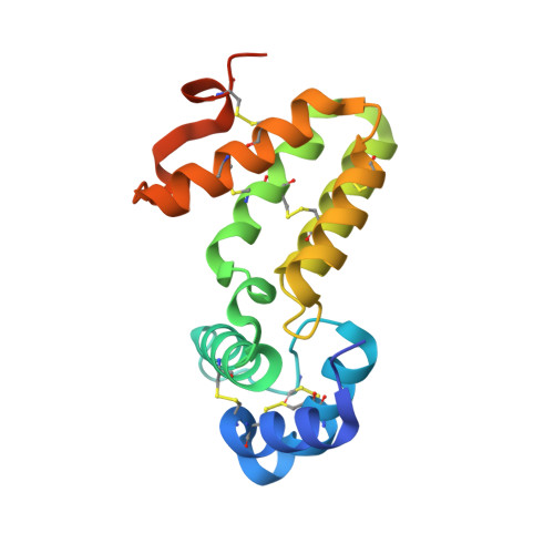
    HER-1 is a secreted protein that promotes male development in the nematode Caenorhabditis elegans. HER-1 inhibits the function of TRA-2A, a multipass integral membrane protein thought to serve as its receptor. We report here the 1.5-A crystal structure of HER-1. The structure was solved by the multiwavelength anomalous diffraction method by using selenomethionyl-substituted HER-1 produced in Chinese hamster ovary cells. The HER-1 structure consists of two all-helical domains and is not closely homologous to any known structure. Sites of amino acid substitutions known to impair HER-1 function were mapped on the HER-1 structure and classified according to the likely mechanism by which they affect HER-1 activity. A subset of these and other amino acid substitutions on the HER-1 surface were assayed for their ability to disrupt interactions between HER-1 and TRA-2A-expressing cells, and a localized region on the HER-1 surface important for mediating this interaction was identified.


  • Organizational Affiliation
    • Department of Biophysics and Biophysical Chemistry and Howard Hughes Medical Institute, The Johns Hopkins University School of Medicine, 725 North Wolfe Street, Baltimore, MD 21205, USA.

Macromolecule Content 

  • Total Structure Weight: 37.09 kDa 
  • Atom Count: 2,564 
  • Modeled Residue Count: 290 
  • Deposited Residue Count: 322 
  • Unique protein chains: 1

Macromolecules

Find similar proteins by:|  3D Structure
Entity ID: 1
MoleculeChains  Sequence LengthOrganismDetailsImage
Her-1 protein
A, B
161Caenorhabditis elegansMutation(s): 2 
Gene Names: HER-1ZK287.8
UniProt
Find proteins for P34704 (Caenorhabditis elegans)
Explore P34704 
Go to UniProtKB:  P34704
Entity Groups
Sequence Clusters30% Identity50% Identity70% Identity90% Identity95% Identity100% Identity
UniProt GroupP34704
Sequence Annotations
Expand
Reference Sequence

Experimental Data & Validation

Experimental Data

  • Method: X-RAY DIFFRACTION
  • Resolution: 1.50 Å
  • R-Value Free:  0.227 (Depositor), 0.227 (DCC) 
  • R-Value Work:  0.209 (Depositor), 0.208 (DCC) 
  • R-Value Observed: 0.209 (Depositor) 
Space Group: P 21 21 21
Unit Cell:
Length ( Å )Angle ( ˚ )
a = 52.97α = 90
b = 58.93β = 90
c = 99γ = 90
Software Package:
Software NamePurpose
CNSrefinement
DENZOdata reduction
SCALEPACKdata scaling
SOLVEphasing

Structure Validation

View Full Validation Report


Entry History 

Deposition Data

Revision History  (Full details and data files)

  • Version 1.0: 2004-08-10
    Type: Initial release
  • Version 1.1: 2008-04-30
    Changes: Version format compliance
  • Version 1.2: 2011-07-13
    Changes: Version format compliance
  • Version 1.3: 2021-10-27
    Changes: Database references, Derived calculations
  • Version 1.4: 2024-10-30
    Changes: Data collection, Structure summary