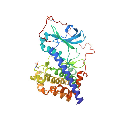
Crystal structure of the E230Q mutant of cAMP-dependent protein kinase reveals an unexpected apoenzyme conformation and an extended N-terminal A helix.
Wu, J., Yang, J., Kannan, N., Madhusudan, N., Xuong, N.H., Ten Eyck, L.F., Taylor, S.S.(2005) Protein Sci 14: 2871-2879
- PubMed: 16253959
- DOI: https://doi.org/10.1110/ps.051715205
- Primary Citation of Related Structures:
1SYK - PubMed Abstract:
Glu230, one of the acidic residues that cluster around the active site of the catalytic subunit of cAMP-dependent protein kinase, plays an important role in substrate recognition. Specifically, its side chain forms a direct salt-bridge interaction with the substrate's P-2 Arg. Previous studies showed that mutation of Glu230 to Gln (E230Q) caused significant decreases not only in substrate binding but also in the rate of phosphoryl transfer. To better understand the importance of Glu230 for structure and function, we solved the crystal structure of the E230Q mutant at 2.8 A resolution. Surprisingly, the mutant preferred an open conformation with no bound ligands observed, even though the crystals were grown in the presence of MgATP and the inhibitor peptide, IP20. This is in contrast to the wild-type protein that, under the same conditions, prefers the closed conformation of a ternary complex. The structure highlights the importance of the electrostatic surface not only for substrate binding and catalysis, but also for the mechanism for closing the active site cleft. This surface mutation clearly disrupts the recognition and binding of substrate peptide so that the enzyme prefers an open conformation that cannot trap ATP. This is consistent with the reinforcing concepts of conformational dynamics and the synergistic binding of ATP and substrate peptide. Another unusual feature of the structure is the observation of the entire N terminus (Gly1-Thr32) assumes an extended alpha-helix conformation. Finally, based on temperature factors, this mutant structure is more stable than the wild-type C-subunit in the apo state.
- Howard Hughes Medical Institute, Department of Chemistry and Biochemistry, University of California-San Diego, 9500 Gilman Drive, Leichtag 415, La Jolla, CA 92093, USA.
Organizational Affiliation:


