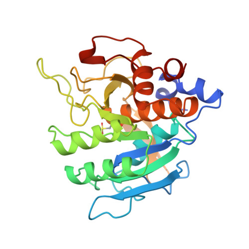
1SUC

CALCIUM-INDEPENDENT SUBTILISIN BY DESIGN


Experimental Data Snapshot

  • Method: X-RAY DIFFRACTION
  • Resolution: 1.80 Å
  • R-Value Observed: 0.177 

wwPDB Validation   3D Report Full Report


This is version 1.5 of the entry. See complete history


Literature

Calcium-independent subtilisin by design.

Gallagher, T.Bryan, P.Gilliland, G.L.

(1993) Proteins 16: 205-213

  • DOI: https://doi.org/10.1002/prot.340160207
  • Primary Citation of Related Structures:  
    1SUB, 1SUC, 1SUD

  • PubMed Abstract: 

    A version of subtilisin BPN' lacking the high affinity calcium site (site A) has been produced through genetic engineering methods, and its crystal structure refined at 1.8 A resolution. This protein and the corresponding version containing the calcium A site are described and compared. The deletion of residues 75-83 was made in the context of four site-specific replacements previously shown to stabilize subtilisin. The helix that in wild type is interrupted by the calcium binding loop, is continuous in the deletion mutant, with normal geometry. A few residues adjacent to the loop, principally those that were involved in calcium coordination, are repositioned and/or destabilized by the deletion. Because refolding is greatly facilitated by the absence of the Ca-loop, this protein offers a new vehicle for analysis and dissection of the folding reaction. This is among the largest internal changes to a protein to be described at atomic resolution.


  • Organizational Affiliation

    Center for Advanced Research in Biotechnology, Maryland Biotechnology Institute, University of Maryland, Shady Grove 20850.


Macromolecules
Find similar proteins by:  (by identity cutoff)  |  3D Structure
Entity ID: 1
MoleculeChains Sequence LengthOrganismDetailsImage
SUBTILISIN BPN' CRB-S3275Bacillus amyloliquefaciensMutation(s): 4 
EC: 3.4.21.62
UniProt
Find proteins for P00782 (Bacillus amyloliquefaciens)
Explore P00782 
Go to UniProtKB:  P00782
Entity Groups  
Sequence Clusters30% Identity50% Identity70% Identity90% Identity95% Identity100% Identity
UniProt GroupP00782
Sequence Annotations
Expand
  • Reference Sequence
Small Molecules
Ligands 2 Unique
IDChains Name / Formula / InChI Key2D Diagram3D Interactions
ACN
Query on ACN

Download Ideal Coordinates CCD File 
C [auth A]ACETONE
C3 H6 O
CSCPPACGZOOCGX-UHFFFAOYSA-N
K
Query on K

Download Ideal Coordinates CCD File 
B [auth A]POTASSIUM ION
K
NPYPAHLBTDXSSS-UHFFFAOYSA-N
Modified Residues  1 Unique
IDChains TypeFormula2D DiagramParent
CSD
Query on CSD
A
L-PEPTIDE LINKINGC3 H7 N O4 SCYS
Experimental Data & Validation

Experimental Data

  • Method: X-RAY DIFFRACTION
  • Resolution: 1.80 Å
  • R-Value Observed: 0.177 
  • Space Group: P 1 21 1
Unit Cell:
Length ( Å )Angle ( ˚ )
a = 41.37α = 90
b = 78.67β = 114.73
c = 36.78γ = 90
Software Package:
Software NamePurpose
PROFFTrefinement

Structure Validation

View Full Validation Report


Entry History 

Deposition Data

Revision History  (Full details and data files)

  • Version 1.0: 1994-01-31
    Type: Initial release
  • Version 1.1: 2008-03-03
    Changes: Version format compliance
  • Version 1.2: 2011-07-13
    Changes: Version format compliance
  • Version 1.3: 2017-11-29
    Changes: Derived calculations, Other
  • Version 1.4: 2021-11-03
    Changes: Database references, Derived calculations
  • Version 1.5: 2024-10-23
    Changes: Data collection, Structure summary