1STR

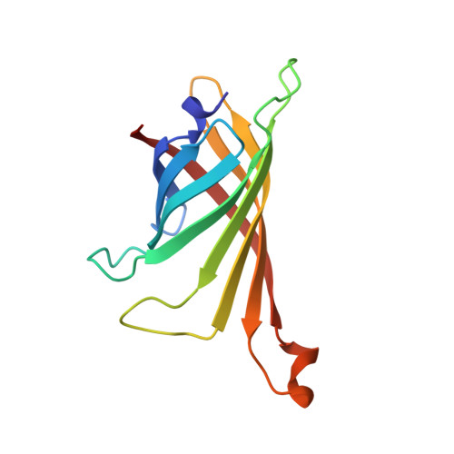
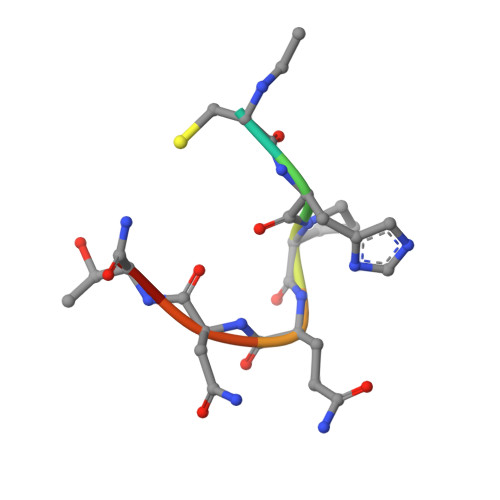
STREPTAVIDIN DIMERIZED BY DISULFIDE-BONDED PEPTIDE AC-CHPQNT-NH2 DIMER


Experimental Data Snapshot

  • Method: X-RAY DIFFRACTION
  • Resolution: 1.80 Å
  • R-Value Free: 0.233 
  • R-Value Work: 0.189 
  • R-Value Observed: 0.189 

wwPDB Validation   3D Report Full Report


This is version 1.4 of the entry. See complete history


Literature

Topochemical catalysis achieved by structure-based ligand design.

Katz, B.A.Cass, R.T.Liu, B.Arze, R.Collins, N.

(1995) J Biol Chem 270: 31210-31218

  • DOI: https://doi.org/10.1074/jbc.270.52.31210
  • Primary Citation of Related Structures:  
    1STR, 1STS

  • PubMed Abstract: 

    Recently, a cyclic peptide ligand, cyclo-Ac-[CHPQG-PPC]-NH2, that binds to streptavidin with high affinity was discovered by screening phage libraries. From the streptavidin-bound crystal structures of cyclo-Ac-[CHPQGPPC]-NH2 and of a related but more weakly binding linear ligand, FSHPQNT, we designed linear thiol-containing streptavidin binding ligands, FCH-PQNT-NH2 and Ac-CHPQNT-NH2, which are dimerized catalytically by the streptavidin crystal lattice of space group I222, as demonstrated by high performance liquid chromatography and mass spectrometry. The catalytic dimerization relies on presentation of the ligand thiols toward one another in the lattice. The streptavidin crystal lattice-mediated catalysis achieved by structure-based design is the first example of catalysis of a chemical reaction by a protein crystal lattice. The spontaneous and crystal catalyzed rates of disulfide formation were determined by high performance liquid chromatography at pH 3.1, 4.0, 5.0, and 6.0. The ratio of the catalyzed to uncatalyzed rate was maximal at pH 3.1 (kcat/kuncat = 3.8), diminishing to 1.2 at pH 6.0. The crystal structures of the streptavidin-bound dimerized peptide ligands, FCHPQNT-NH2 dimer at 1.95 A and Ac-CHPQNT-NH2 dimer at 1.80 A, are described and compared with the structures of streptavidin-bound FSHPQNT monomer and cyclo-Ac-[CHPQGPPC]-NH2 dimer.


  • Organizational Affiliation

    Arris Pharmaceutical Corporation, South San Francisco, California 94080, USA.


Macromolecules
Find similar proteins by:  (by identity cutoff)  |  3D Structure
Entity ID: 1
MoleculeChains Sequence LengthOrganismDetailsImage
STREPTAVIDINA [auth B],
B [auth D]
123Streptomyces avidiniiMutation(s): 0 
UniProt
Find proteins for P22629 (Streptomyces avidinii)
Explore P22629 
Go to UniProtKB:  P22629
Entity Groups  
Sequence Clusters30% Identity50% Identity70% Identity90% Identity95% Identity100% Identity
UniProt GroupP22629
Sequence Annotations
Expand
  • Reference Sequence

Find similar proteins by:  Sequence   |   3D Structure  

Entity ID: 2
MoleculeChains Sequence LengthOrganismDetailsImage
AC-CHPQNT-NH2C [auth M],
D [auth P]
8N/AMutation(s): 0 
Sequence Annotations
Expand
  • Reference Sequence
Experimental Data & Validation

Experimental Data

  • Method: X-RAY DIFFRACTION
  • Resolution: 1.80 Å
  • R-Value Free: 0.233 
  • R-Value Work: 0.189 
  • R-Value Observed: 0.189 
  • Space Group: I 2 2 2
Unit Cell:
Length ( Å )Angle ( ˚ )
a = 96.74α = 90
b = 106.04β = 90
c = 48.39γ = 90
Software Package:
Software NamePurpose
X-PLORmodel building
X-PLORrefinement
SADIEdata reduction
SAINTdata reduction
X-PLORphasing

Structure Validation

View Full Validation Report


Entry History 

Deposition Data

Revision History  (Full details and data files)

  • Version 1.0: 1996-03-08
    Type: Initial release
  • Version 1.1: 2008-03-24
    Changes: Version format compliance
  • Version 1.2: 2011-07-13
    Changes: Version format compliance
  • Version 1.3: 2024-06-05
    Changes: Data collection, Database references, Derived calculations, Other
  • Version 1.4: 2024-11-13
    Changes: Structure summary