1SQW | pdb_00001sqw

Crystal structure of KD93, a novel protein expressed in the human pro


Experimental Data Snapshot

  • Method: X-RAY DIFFRACTION
  • Resolution: 1.90 Å
  • R-Value Free: 
    0.261 (Depositor), 0.258 (DCC) 
  • R-Value Work: 
    0.225 (Depositor), 0.217 (DCC) 

wwPDB Validation 3D Report Full Report

Validation slider image for 1SQW

This is version 1.3 of the entry. See complete history

Literature

Crystal structure of KD93, a novel protein expressed in human hematopoietic stem/progenitor cells.

Liu, J.F.Wang, X.Q.Wang, Z.X.Chen, J.R.Jiang, T.An, X.M.Chang, W.R.Liang, D.C.

(2004) J Struct Biol 148: 370-374

  • DOI: https://doi.org/10.1016/j.jsb.2004.06.010
  • Primary Citation Related Structures: 
    1SQW

  • PubMed Abstract: 

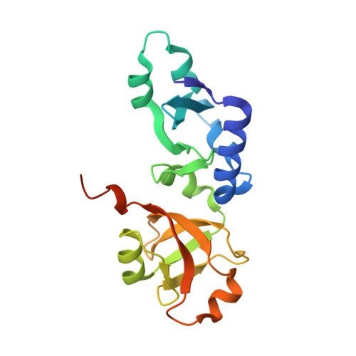
    The crystal structure of a novel hypothetical protein, KD93, expressed in human hematopoietic stem/progenitor cells, was determined at 1.9A resolution using the multiple-wavelength anomalous dispersion (MAD) method. The protein KD93, which is encoded by the open reading frame HSPC031, is a NIP7 homologue and belongs to the UPF0113 family. The structural and functional information for the group of homologues has not yet been determined. Crystallographic analysis revealed that the overall fold of KD93 consists of two interlinked alpha/beta domains. Structure-based homology analysis with DALI revealed that the C domain of KD93 matches the PUA domain of some RNA modification enzymes, especially that of archaeosine tRNA-ribosyltransferase (ArcTGT), which suggests that its possible molecular function is related to RNA binding. The difference between the RNA binding regions of KD93 and ArcTGT in amino acid constitution and surface electrostatic potential indicate that they may have different RNA binding modes. The N domain of KD93 is a unique structure with no obvious similarity to other proteins with known three-dimensional structures. The high-resolution structure of KD93 provides a first view of a member of the family of hypothetical proteins. And the structure provides a framework to deduce and assay the molecular function of other proteins of the UPF0113 family.


  • Organizational Affiliation
    • National Laboratory of Biomacromolecules, Institute of Biophysics, Academia Sinica, Beijing 100101, People's Republic of China.

Macromolecule Content 

  • Total Structure Weight: 21.56 kDa 
  • Atom Count: 1,540 
  • Modeled Residue Count: 176 
  • Deposited Residue Count: 188 
  • Unique protein chains: 1

Macromolecules

Find similar proteins by:|  3D Structure
Entity ID: 1
MoleculeChains  Sequence LengthOrganismDetailsImage
Saccharomyces cerevisiae Nip7p homolog188Homo sapiensMutation(s): 0 
Gene Names: novel gene
UniProt & NIH Common Fund Data Resources
Find proteins for Q9Y221 (Homo sapiens)
Explore Q9Y221 
Go to UniProtKB:  Q9Y221
PHAROS:  Q9Y221
GTEx:  ENSG00000132603 
Entity Groups
Sequence Clusters30% Identity50% Identity70% Identity90% Identity95% Identity100% Identity
UniProt GroupQ9Y221
Sequence Annotations
Expand
Reference Sequence

Experimental Data & Validation

Experimental Data

  • Method: X-RAY DIFFRACTION
  • Resolution: 1.90 Å
  • R-Value Free:  0.261 (Depositor), 0.258 (DCC) 
  • R-Value Work:  0.225 (Depositor), 0.217 (DCC) 
Space Group: P 41 21 2
Unit Cell:
Length ( Å )Angle ( ˚ )
a = 57.203α = 90
b = 57.203β = 90
c = 132.048γ = 90
Software Package:
Software NamePurpose
DENZOdata reduction
SCALEPACKdata scaling
SOLVEphasing
CNSrefinement

Structure Validation

View Full Validation Report


Entry History 

Deposition Data

Revision History  (Full details and data files)

  • Version 1.0: 2005-03-22
    Type: Initial release
  • Version 1.1: 2008-04-29
    Changes: Version format compliance
  • Version 1.2: 2011-07-13
    Changes: Version format compliance
  • Version 1.3: 2024-02-14
    Changes: Data collection, Database references