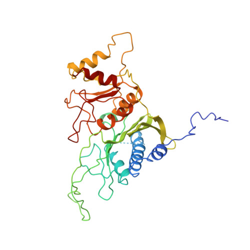
Crystal structure of spinach chloroplast fructose-1,6-bisphosphatase at 2.8 A resolution.
Villeret, V., Huang, S., Zhang, Y., Xue, Y., Lipscomb, W.N.(1995) Biochemistry 34: 4299-4306
- PubMed: 7703243
- DOI: https://doi.org/10.1021/bi00013a019
- Primary Citation of Related Structures:
1SPI - PubMed Abstract:
The three-dimensional structure of the spinach chloroplast fructose-1,6-bisphosphatase (Fru-1,6-Pase) has been solved by the molecular replacement method at 2.8 A resolution and refined to a crystallographic R factor of 0.203. The enzyme is composed of four monomers and displays pseudo D2 symmetry. Comparison with the allosteric Fru-1,6-Pase from pig kidney shows orientationally displaced dimers within the quaternary structure of the chloroplast enzyme. When the C1C2 dimers of the two enzymes are superimposed, the C3C4 dimer of the chloroplast enzyme is rotated 20 degrees and 5 degrees relative to the C3C4 dimer of the R and T forms of the pig kidney enzyme, respectively. This new quaternary structure, designated as S, may be described as a super-T form and is outside of the pathway of the allosteric transition which occurs in the pig kidney enzyme, which shows a 15 degrees rotation between T and R forms. Chloroplast Fru-1,6-Pase, unlike the pig kidney enzyme, is insensitive to allosteric transformation by AMP. Structural changes in the AMP binding site involving mainly helices H1, H2, and H3 and the loop between H1 and H2 at the dimer interface interfere with binding of the phosphate of AMP. Finally, the location of cysteines residues provides a basis for a preliminary discussion of the activation of the enzyme by reduction of cysteines via the ferredoxin-thioredoxin f system; this process is complementary to activation by pH changes, Mg2+ or Ca2+, Fru-1,6-P2, and possibly Fru-2,6-P2.
- Gibbs Chemical Laboratory, Harvard University, Cambridge, Massachusetts 02138, USA.
Organizational Affiliation:


