1SPG | pdb_00001spg

CARBONMONOXY HEMOGLOBIN FROM THE TELEOST FISH LEIOSTOMUS XANTHURUS


Experimental Data Snapshot

  • Method: X-RAY DIFFRACTION
  • Resolution: 1.95 Å
  • R-Value Free: 
    0.245 (Depositor) 
  • R-Value Work: 
    0.191 (Depositor) 
  • R-Value Observed: 
    0.191 (Depositor) 

wwPDB Validation 3D Report Full Report

Validation slider image for 1SPG

This is version 1.4 of the entry. See complete history

Literature

Structural basis for the root effect in haemoglobin.

Mylvaganam, S.E.Bonaventura, C.Bonaventura, J.Getzoff, E.D.

(1996) Nat Struct Biol 3: 275-283

  • DOI: https://doi.org/10.1038/nsb0396-275
  • Primary Citation Related Structures: 
    1SPG

  • PubMed Abstract: 

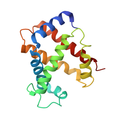
    The remarkable ability of root effect haemoglobins to pump oxygen against high O2 gradients results from extreme, acid-induced reductions in O2 affinity and cooperativity. The long-sought mechanism for the root effect, revealed by the 2 angstrom crystal structure of the ligand-bound haemoglobin from Leiostomus xanthurus at pH 7.5, unexpectedly involves modulation of the R-state. Key residues strategically assemble positive-charge clusters across the allosteric beta1 beta2-interface in the R-state. At low pH, protonation of the beta N terminus and His 147(HC3)beta within these clusters is postulated to destabilize the R-state and promote the acid-triggered, allosteric R-->T switch with concomitant O2 release. Surprisingly, a set of residues specific to root effect haemoglobins recruit additional residues, conserved among most haemoglobins, to produce the root effect.


  • Organizational Affiliation
    • The Scripps Research Institute, La Jolla, California 92037, USA.

Macromolecule Content 

  • Total Structure Weight: 33.29 kDa 
  • Atom Count: 2,518 
  • Modeled Residue Count: 291 
  • Deposited Residue Count: 291 
  • Unique protein chains: 2

Macromolecules

Find similar proteins by:|  3D Structure
Entity ID: 1
MoleculeChains  Sequence LengthOrganismDetailsImage
HEMOGLOBIN144Leiostomus xanthurusMutation(s): 0 
UniProt
Find proteins for P56250 (Leiostomus xanthurus)
Explore P56250 
Go to UniProtKB:  P56250
Entity Groups
Sequence Clusters30% Identity50% Identity70% Identity90% Identity95% Identity100% Identity
UniProt GroupP56250
Sequence Annotations
Expand
Reference Sequence
Find similar proteins by:|  3D Structure
Entity ID: 2
MoleculeChains  Sequence LengthOrganismDetailsImage
HEMOGLOBIN147Leiostomus xanthurusMutation(s): 0 
UniProt
Find proteins for P56251 (Leiostomus xanthurus)
Explore P56251 
Go to UniProtKB:  P56251
Entity Groups
Sequence Clusters30% Identity50% Identity70% Identity90% Identity95% Identity100% Identity
UniProt GroupP56251
Sequence Annotations
Expand
Reference Sequence

Experimental Data & Validation

Experimental Data

  • Method: X-RAY DIFFRACTION
  • Resolution: 1.95 Å
  • R-Value Free:  0.245 (Depositor) 
  • R-Value Work:  0.191 (Depositor) 
  • R-Value Observed: 0.191 (Depositor) 
Space Group: C 1 2 1
Unit Cell:
Length ( Å )Angle ( ˚ )
a = 89.6α = 90
b = 75.6β = 141.9
c = 69.7γ = 90
Software Package:
Software NamePurpose
X-PLORmodel building
X-PLORrefinement
XENGENdata reduction
XENGENdata scaling
X-PLORphasing

Structure Validation

View Full Validation Report


Entry History 

Deposition Data

Revision History  (Full details and data files)

  • Version 1.0: 1997-03-12
    Type: Initial release
  • Version 1.1: 2008-03-24
    Changes: Version format compliance
  • Version 1.2: 2011-07-13
    Changes: Derived calculations, Version format compliance
  • Version 1.3: 2024-06-05
    Changes: Data collection, Database references, Derived calculations, Other
  • Version 1.4: 2024-10-16
    Changes: Structure summary