1SOO

ADENYLOSUCCINATE SYNTHETASE INHIBITED BY HYDANTOCIDIN 5'-MONOPHOSPHATE


Experimental Data Snapshot

  • Method: X-RAY DIFFRACTION
  • Resolution: 2.60 Å
  • R-Value Work: 0.187 
  • R-Value Observed: 0.187 

Starting Model: experimental
View more details

wwPDB Validation   3D Report Full Report


Ligand Structure Quality Assessment 


This is version 1.4 of the entry. See complete history


Literature

The mode of action and the structure of a herbicide in complex with its target: binding of activated hydantocidin to the feedback regulation site of adenylosuccinate synthetase.

Fonne-Pfister, R.Chemla, P.Ward, E.Girardet, M.Kreuz, K.E.Honzatko, R.B.Fromm, H.J.Schar, H.P.Grutter, M.G.Cowan-Jacob, S.W.

(1996) Proc Natl Acad Sci U S A 93: 9431-9436

  • DOI: https://doi.org/10.1073/pnas.93.18.9431
  • Primary Citation of Related Structures:  
    1SON, 1SOO

  • PubMed Abstract: 

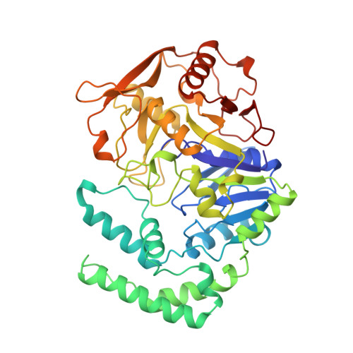
    (+)-Hydantocidin, a recently discovered natural spironucleoside with potent herbicidal activity, is shown to be a proherbicide that, after phosphorylation at the 5' position, inhibits adenylosuccinate synthetase, an enzyme involved in de novo purine synthesis. The mode of binding of hydantocidin 5'-monophosphate to the target enzyme was analyzed by determining the crystal structure of the enzyme-inhibitor complex at 2.6-A resolution. It was found that adenylosuccinate synthetase binds the phosphorylated compound in the same fashion as it does adenosine 5'-monophosphate, the natural feedback regulator of this enzyme. This work provides the first crystal structure of a herbicide-target complex reported to date.


  • Organizational Affiliation

    Pharmaceutical Division, Ciba-Geigy Ltd., Basel, Switzerland.


Macromolecules
Find similar proteins by:  (by identity cutoff)  |  3D Structure
Entity ID: 1
MoleculeChains Sequence LengthOrganismDetailsImage
ADENYLOSUCCINATE SYNTHETASE431Escherichia coliMutation(s): 0 
EC: 6.3.4.4
UniProt
Find proteins for P0A7D4 (Escherichia coli (strain K12))
Explore P0A7D4 
Go to UniProtKB:  P0A7D4
Entity Groups  
Sequence Clusters30% Identity50% Identity70% Identity90% Identity95% Identity100% Identity
UniProt GroupP0A7D4
Sequence Annotations
Expand
  • Reference Sequence
Experimental Data & Validation

Experimental Data

  • Method: X-RAY DIFFRACTION
  • Resolution: 2.60 Å
  • R-Value Work: 0.187 
  • R-Value Observed: 0.187 
  • Space Group: P 43 21 2
Unit Cell:
Length ( Å )Angle ( ˚ )
a = 70.5α = 90
b = 70.5β = 90
c = 198.8γ = 90
Software Package:
Software NamePurpose
X-PLORmodel building
X-PLORrefinement
X-PLORphasing

Structure Validation

View Full Validation Report


Ligand Structure Quality Assessment 


Entry History 

Deposition Data

Revision History  (Full details and data files)

  • Version 1.0: 1997-09-04
    Type: Initial release
  • Version 1.1: 2008-03-24
    Changes: Version format compliance
  • Version 1.2: 2011-07-13
    Changes: Derived calculations, Version format compliance
  • Version 1.3: 2018-04-18
    Changes: Data collection, Other
  • Version 1.4: 2023-08-09
    Changes: Database references, Derived calculations, Refinement description