1SNM | pdb_00001snm

ACTIVE SITE MUTANT GLU-43 (RIGHT ARROW) ASP IN STAPHYLOCOCCAL NUCLEASE DISPLAYS NONLOCAL STRUCTURAL CHANGES


Experimental Data Snapshot

  • Method: X-RAY DIFFRACTION
  • Resolution: 1.74 Å
  • R-Value Work: 
    0.175 (DCC) 
  • R-Value Observed: 
    0.174 (Depositor) 

wwPDB Validation 3D Report Full Report

Validation slider image for 1SNM

Ligand Structure Quality Assessment 


This is version 1.4 of the entry. See complete history

Literature

Active site mutant Glu-43 --> Asp in staphylococcal nuclease displays nonlocal structural changes.

Loll, P.J.Lattman, E.E.

(1990) Biochemistry 29: 6866-6873

  • DOI: https://doi.org/10.1021/bi00481a016
  • Primary Citation Related Structures: 
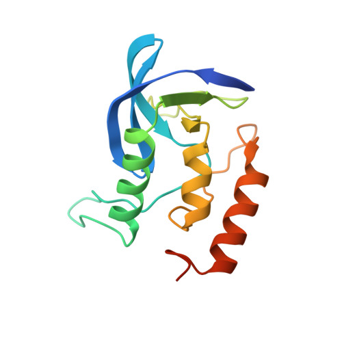
    1SNM

  • PubMed Abstract: 

    The crystal structure of the Glu-43----Asp mutant of staphylococcal nuclease complexed with Ca2+ and the inhibitor thymidine 3',5'-bisphosphate (pdTp) has been determined and refined by restrained least-squares methods to a conventional crystallographic R value of 0.174 at a resolution of 1.74 A. Throughout most of the structure, the conformation of the backbone atoms of the mutant is similar to that of the wild-type protein; however, the seemingly conservative mutation Glu----Asp has significantly perturbed the structure of a loop adjacent to the active site, as well as giving rise to looser binding of the essential calcium ion and to a less extensive network of bound water molecules in the active site. Crystal contacts that extend into the active site have also been altered by this amino acid substitution. The changes caused by this mutation are considerably more drastic than would have been predicted and should serve as caveats to those who would draw conclusions about structure-function relationships on the basis of site-directed mutagenesis experiments in the absence of structural data.


  • Organizational Affiliation
    • Department of Biochemistry and Molecular Biology, University of Chicago, Illinois 60637.

Macromolecule Content 

  • Total Structure Weight: 17.27 kDa 
  • Atom Count: 1,168 
  • Modeled Residue Count: 136 
  • Deposited Residue Count: 149 
  • Unique protein chains: 1

Macromolecules

Find similar proteins by:|  3D Structure
Entity ID: 1
MoleculeChains  Sequence LengthOrganismDetailsImage
THERMONUCLEASE PRECURSOR149Staphylococcus aureusMutation(s): 1 
EC: 3.1.31.1
UniProt
Find proteins for P00644 (Staphylococcus aureus)
Explore P00644 
Go to UniProtKB:  P00644
Entity Groups
Sequence Clusters30% Identity50% Identity70% Identity90% Identity95% Identity100% Identity
UniProt GroupP00644
Sequence Annotations
Expand
Reference Sequence

Small Molecules

Experimental Data & Validation

Experimental Data

  • Method: X-RAY DIFFRACTION
  • Resolution: 1.74 Å
  • R-Value Work:  0.175 (DCC) 
  • R-Value Observed: 0.174 (Depositor) 
Space Group: P 41
Unit Cell:
Length ( Å )Angle ( ˚ )
a = 48.59α = 90
b = 48.59β = 90
c = 63.38γ = 90
Software Package:
Software NamePurpose
PROLSQrefinement

Structure Validation

View Full Validation Report


Ligand Structure Quality Assessment 


Entry History 

Deposition Data

Revision History  (Full details and data files)

  • Version 1.0: 1991-07-15
    Type: Initial release
  • Version 1.1: 2008-03-03
    Changes: Version format compliance
  • Version 1.2: 2011-07-13
    Changes: Version format compliance
  • Version 1.3: 2017-11-29
    Changes: Derived calculations, Other
  • Version 1.4: 2024-02-14
    Changes: Data collection, Database references, Derived calculations