1SM8 | pdb_00001sm8

M. tuberculosis dUTPase complexed with chromium and dUTP


Experimental Data Snapshot

  • Method: X-RAY DIFFRACTION
  • Resolution: 2.90 Å
  • R-Value Free: 
    0.282 (Depositor), 0.276 (DCC) 
  • R-Value Work: 
    0.235 (Depositor), 0.233 (DCC) 
  • R-Value Observed: 
    0.235 (Depositor) 

Starting Model: experimental
View more details

wwPDB Validation 3D Report Full Report

Validation slider image for 1SM8

Ligand Structure Quality Assessment 


This is version 1.3 of the entry. See complete history

Literature

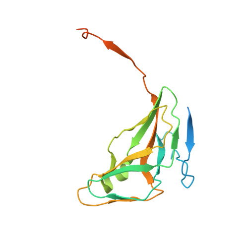
Crystal structure of the Mycobacterium tuberculosis dUTPase: insights into the catalytic mechanism.

Chan, S.Segelke, B.Lekin, T.Krupka, H.Cho, U.S.Kim, M.-Y.So, M.Kim, C.-Y.Naranjo, C.M.Rogers, Y.C.Park, M.S.Waldo, G.S.Pashkov, I.Cascio, D.Perry, J.L.Sawaya, M.R.

(2004) J Mol Biology 341: 503-517

  • DOI: https://doi.org/10.1016/j.jmb.2004.06.028
  • Primary Citation Related Structures: 
    1MQ7, 1SIX, 1SJN, 1SLH, 1SM8, 1SMC, 1SNF

  • PubMed Abstract: 

    The structure of Mycobacterium tuberculosis dUTP nucleotidohydrolase (dUTPase) has been determined at 1.3 Angstrom resolution in complex with magnesium ion and the non-hydrolyzable substrate analog, alpha,beta-imido dUTP. dUTPase is an enzyme essential for depleting potentially toxic concentrations of dUTP in the cell. Given the importance of its biological role, it has been proposed that inhibiting M.tuberculosis dUTPase might be an effective means to treat tuberculosis infection in humans. The crystal structure presented here offers some insight into the potential for designing a specific inhibitor of the M.tuberculosis dUTPase enzyme. The structure also offers new insights into the mechanism of dUTP hydrolysis by providing an accurate representation of the enzyme-substrate complex in which both the metal ion and dUTP analog are included. The structure suggests that inclusion of a magnesium ion is important for stabilizing the position of the alpha-phosphorus for an in-line nucleophilic attack. In the absence of magnesium, the alpha-phosphate of dUTP can have either of the two positions which differ by 4.5 Angstrom. A transiently ordered C-terminal loop further assists catalysis by shielding the general base, Asp83, from solvent thus elevating its pK(a) so that it might in turn activate a tightly bound water molecule for nucleophilic attack. The metal ion coordinates alpha, beta, and gamma phosphate groups with tridentate geometry identical with that observed in the crystal structure of DNA polymerase beta complexed with magnesium and dNTP analog, revealing some common features in catalytic mechanism.


  • Organizational Affiliation
    • UCLA-DOE Laboratory of Structural Biology and Molecular Medicine, 206 Boyer Hall, Box 951570, Los Angeles, CA 90095-1570, USA.

Macromolecule Content 

  • Total Structure Weight: 55.72 kDa 
  • Atom Count: 3,028 
  • Modeled Residue Count: 397 
  • Deposited Residue Count: 522 
  • Unique protein chains: 1

Macromolecules

Find similar proteins by:|  3D Structure
Entity ID: 1
MoleculeChains  Sequence LengthOrganismDetailsImage
Deoxyuridine 5'-triphosphate nucleotidohydrolase
A, B, C
174Mycobacterium tuberculosisMutation(s): 0 
Gene Names: DUTRV2697CMT2771MTCY05A6.18CMB2716C
EC: 3.6.1.23
UniProt
Find proteins for P9WNS5 (Mycobacterium tuberculosis (strain ATCC 25618 / H37Rv))
Explore P9WNS5 
Go to UniProtKB:  P9WNS5
Entity Groups
Sequence Clusters30% Identity50% Identity70% Identity90% Identity95% Identity100% Identity
UniProt GroupP9WNS5
Sequence Annotations
Expand
Reference Sequence

Small Molecules

Ligands 4 Unique
IDChains Name / Formula / InChI Key2D Diagram3D Interactions
DUT

Query on DUT


Download:Ideal Coordinates CCD File
G [auth B],
J [auth C],
K [auth C]
DEOXYURIDINE-5'-TRIPHOSPHATE
C9 H15 N2 O14 P3
AHCYMLUZIRLXAA-SHYZEUOFSA-N
TRS

Query on TRS


Download:Ideal Coordinates CCD File
H [auth B]2-AMINO-2-HYDROXYMETHYL-PROPANE-1,3-DIOL
C4 H12 N O3
LENZDBCJOHFCAS-UHFFFAOYSA-O
NO3

Query on NO3


Download:Ideal Coordinates CCD File
E [auth A]NITRATE ION
N O3
NHNBFGGVMKEFGY-UHFFFAOYSA-N
CR

Query on CR


Download:Ideal Coordinates CCD File
D [auth A],
F [auth B],
I [auth C]
CHROMIUM ION
Cr
BFGKITSFLPAWGI-UHFFFAOYSA-N

Experimental Data & Validation

Experimental Data

  • Method: X-RAY DIFFRACTION
  • Resolution: 2.90 Å
  • R-Value Free:  0.282 (Depositor), 0.276 (DCC) 
  • R-Value Work:  0.235 (Depositor), 0.233 (DCC) 
  • R-Value Observed: 0.235 (Depositor) 
Space Group: P 21 21 21
Unit Cell:
Length ( Å )Angle ( ˚ )
a = 57.934α = 90
b = 77.714β = 90
c = 91.619γ = 90
Software Package:
Software NamePurpose
CNSrefinement
DENZOdata reduction
SCALEPACKdata scaling
EPMRphasing

Structure Validation

View Full Validation Report


Ligand Structure Quality Assessment 


Entry History 

Revision History  (Full details and data files)

  • Version 1.0: 2004-03-16
    Type: Initial release
  • Version 1.1: 2008-04-29
    Changes: Version format compliance
  • Version 1.2: 2011-07-13
    Changes: Version format compliance
  • Version 1.3: 2023-08-23
    Changes: Data collection, Database references, Derived calculations, Refinement description